. 2020 May 19;86(11):e02920-19.
doi: 10.1128/AEM.02920-19. Print 2020 May 19.

Synergistic Cues from Diverse Bacteria Enhance Multicellular Development in a Choanoflagellate

Affiliations

Synergistic Cues from Diverse Bacteria Enhance Multicellular Development in a Choanoflagellate

Ella V Ireland et al. Appl Environ Microbiol. .

Abstract

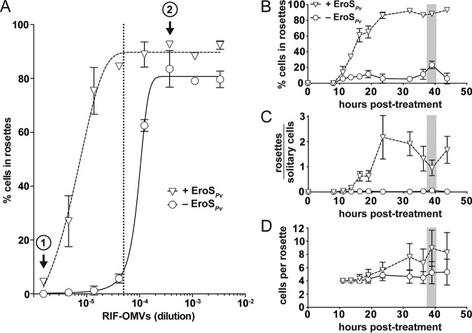
Bacteria regulate the life histories of diverse eukaryotes, but relatively little is known about how eukaryotes interpret and respond to multiple bacterial cues encountered simultaneously. To explore how a eukaryote might respond to a combination of bioactive molecules from multiple bacteria, we treated the choanoflagellate Salpingoeca rosetta with two sets of bacterial cues, one that induces mating and another that induces multicellular development. We found that simultaneous exposure to both sets of cues enhanced multicellular development in S. rosetta, eliciting both larger multicellular colonies and an increase in the number of colonies. Thus, rather than conveying conflicting sets of information, these distinct bacterial cues synergize to augment multicellular development. This study demonstrates how a eukaryote can integrate and modulate its response to cues from diverse bacteria, underscoring the potential impact of complex microbial communities on eukaryotic life histories.IMPORTANCE Eukaryotic biology is profoundly influenced by interactions with diverse environmental and host-associated bacteria. However, it is not well understood how eukaryotes interpret multiple bacterial cues encountered simultaneously. This question has been challenging to address because of the complexity of many eukaryotic model systems and their associated bacterial communities. Here, we studied a close relative of animals, the choanoflagellate Salpingoeca rosetta, to explore how eukaryotes respond to diverse bacterial cues. We found that a bacterial chondroitinase that induces mating on its own can also synergize with bacterial lipids that induce multicellular "rosette" development. When encountered together, these cues enhance rosette development, resulting in both the formation of larger rosettes and an increase in the number of rosettes compared to rosette development in the absence of the chondroitinase. These findings highlight how synergistic interactions among bacterial cues can influence the biology of eukaryotes.

Keywords: EroS; RIF-1; Salpingoeca rosetta; choanoflagellate; chondroitinase; host microbe; multicellularity; outer membrane vesicles; rosette-inducing factor; sulfonolipid.


Figures

FIG 1
FIG 1
Rosettes swarm in response to the EroSPv mating factor. (A to C) Bacterial cues regulate mating and multicellularity in S. rosetta. Bars, 10 μm. (A) S. rosetta grown in the presence of the prey bacterium E. pacifica (Ctrl) proliferated as solitary cells. This culture served as the foundation for all experiments in this study. A typical S. rosetta cell has an apical collar (arrowhead) surrounding a single flagellum (arrow). (B) S. rosetta formed mating swarms within 0.5 h of treatment with the bacterially produced chondroitinase EroSPv. (C) S. rosetta solitary cells developed into rosettes through serial rounds of cell division within 24 h of treatment with RIF-OMVs from the bacterium A. machipongonensis. (D and E) Rosettes swarm in the presence, but not in the absence, of EroSPv. Bars, 50 μm. (D) After 24 h of treatment with a 1:1,000 dilution of RIF-OMVs and BSA (carrier control), solitary cells in an SrEpac culture developed into rosettes (arrowheads) but did not swarm. (E) Swarms of rosettes (arrows) formed after 24 h of treatment with a 1:1,000 dilution of RIF-OMVs followed by 0.5 h of treatment with 0.05 U/ml (0.2 to 1 μg/ml, ∼2 to 8 nM) EroSPv. (F) Scatterplot of the surface areas of cell clusters from SrEpac cultures treated with a 1:1,000 dilution of RIF-OMVs for 24 h followed by 0.5 h of incubation either with BSA (carrier control) or with 0.05 U/ml (0.2 to 1 μg/ml, ∼2 to 8 nM) EroSPv. According to the approach described in reference , we generated a binary mask to measure cell cluster area (the area of each cell, rosette, or swarm) (see Fig. S1 in the supplemental material). EroSPv treatment resulted in clusters of cells, including swarms of rosettes (median, 58.7 μm2; interquartile range, 21.6 to 98.0 μm2), whose areas were significantly larger than those measured in the rosette-only control (median, 35.5 μm2; interquartile range, 17.8 to 65.9 μm2) (Kolmogorov-Smirnov test). In total, 875 cell cluster areas from 3 biological replicates were plotted for the cultures treated with RIF-OMVs, and 1,359 cell cluster areas from 3 biological replicates were plotted for the cultures treated with RIF-OMVs plus EroSPv.
FIG 2
FIG 2
The mating inducer EroSPv enhances rosette development. (A) EroSPv enhances rosette induction by RIF-OMVs. Treatment of SrEpac with increasing concentrations of RIF-OMVs (circles) and BSA (carrier control) resulted in a concomitant increase in the percentage of cells in rosettes. Cotreatment of SrEpac with RIF-OMVs and 0.05 U/ml (0.2 to 1 μg/ml, ∼2 to 8 nM) EroSPv (triangles) resulted in rosette development at concentrations of RIF-OMVs that did not otherwise induce rosettes (e.g., at circle 1). EroSPv also increased the maximum percentage of cells in rosettes at saturating concentrations of RIF-OMVs (e.g., at circle 2). The 1:20,000 dilution of RIF-OMVs used for the sensitized rosette induction assays in panels B to D is indicated with a vertical dotted line. (B) Cotreatment of SrEpac with 0.05 U/ml (0.2 to 1 μg/ml, ∼2 to 8 nM) EroSPv and a 1:20,000 dilution of RIF-OMVs leads to a dramatic increase in percentage of cells in rosettes throughout the course of rosette development relative to that in SrEpac treated only with RIF-OMVs and BSA (carrier control). After 39 h (shaded bar) of cotreatment with RIF-OMVs and EroSPv (triangles), 88.2% ± 2.7% of S. rosetta cells were in rosettes, compared with 23.4% ± 4.9% of cells treated with RIF-OMVs alone (circles). (C) EroSPv increased the ratio of rosettes to solitary cells in SrEpac cultures treated with RIF-OMVs. After 39 h (shaded bar) of cotreatment with a 1:20,000 dilution of RIF-OMVs and 0.05 U/ml (0.2 to 1 μg/ml, ∼2 to 8 nM) EroSPv (triangles), the ratio of rosettes to solitary cells was 0.96 ± 0.31 compared with 0.06 ± 0.02 after treatment with RIF-OMVs and BSA (carrier control) (circles). (D) EroSPv increased the number of cells per rosette in RIF-OMV-treated SrEpac cultures. After 39 h (shaded bar) of cotreatment with a 1:20,000 dilution of RIF-OMVs and 0.05 U/ml (0.2 to 1 μg/ml, ∼2 to 8 nM) EroSPv (triangles), there were 8.9 ± 2.7 S. rosetta cells per rosette colony compared with 5.3 ± 1.7 cells per rosette colony after treatment with RIF-OMVs alone and BSA (carrier control) (circles). Means ± SDs from 3 biological replicates are plotted for each panel (A to D).
FIG 3
FIG 3
Purified RIFs and EroS are sufficient for enhancement of rosette induction. (A) Cotreatment of SrEpac with 10 μg/ml (16.7 μM) HPLC-purified RIFs and 0.05 U/ml (0.2 to 1 μg/ml, ∼2 to 8 nM) EroSPv for 24 h resulted in an increase in the percentage of S. rosetta cells in rosettes compared to that after treatment with HPLC-purified RIFs and BSA (carrier control). Rosettes do not form in the absence of RIFs. Means ± SDs from 2 biological replicates are plotted (unpaired t test). (B) Cotreatment of SrEpac with a 1:20,000 dilution of RIF-OMVs and either 0.1% EroS from V. fischeri (EroSVf) or 0.05 U/ml (0.2 to 1 μg/ml, ∼2 to 8 nM) EroS from P. vulgaris (EroSPv) for 24 h resulted in an increase in the percentage of rosette colonies compared to that after treatment with RIF-OMVs and BSA (carrier control). Means ± SDs from 5 biological replicates (RIF-OMVs alone, RIF-OMVs plus EroSPv) or 4 biological replicates (RIF-OMVs plus EroSVf) are plotted (unpaired t test).
FIG 4
FIG 4
S. rosetta integration of bacterial cues. S. rosetta phenotypes induced over time by EroSPv, RIF-OMVs, and the synergistic effect of both cues. (A) Untreated SrEpac proliferates as solitary cells. (B) Treatment with EroSPv induces swarming of unrelated cells within 0.5 h. Cells with different genotypes (depicted as cells of different colors) can gather together to form nonclonal swarms. (C) Treatment with RIF-OMVs induces rosette development through cell division within 11 to 24 h. Cells in rosettes arise through serial rounds of cell division and share the same genotype (depicted as cells of the same color). (D) Cotreatment with RIF-OMVs and EroSPv for 0.5 h results in swarming, showing that RIF-OMVs do not interfere with or enhance the activity of EroSPv. (E) After 11 to 24 h of cotreatment with RIF-OMVs and EroSPv, rosettes develop and swarm. Compared to that after treatment with RIF-OMVs alone, cotreatment with RIF-OMVs and EroSPv induces the development of more rosettes and rosettes containing more cells.

References

    1. McFall-Ngai MJ. 2002. Unseen forces: the influence of bacteria on animal development. Dev Biol 242:1–14. doi: 10.1006/dbio.2001.0522. - DOI - PubMed
    1. Woznica A, King N. 2018. Lessons from simple marine models on the bacterial regulation of eukaryotic development. Curr Opin Microbiol 43:108–116. doi: 10.1016/j.mib.2017.12.013. - DOI - PMC - PubMed
    1. McFall-Ngai M, Hadfield MG, Bosch TCG, Carey HV, Domazet-Lošo T, Douglas AE, Dubilier N, Eberl G, Fukami T, Gilbert SF, Hentschel U, King N, Kjelleberg S, Knoll AH, Kremer N, Mazmanian SK, Metcalf JL, Nealson K, Pierce NE, Rawls JF, Reid A, Ruby EG, Rumpho M, Sanders JG, Tautz D, Wernegreen JJ. 2013. Animals in a bacterial world, a new imperative for the life sciences. Proc Natl Acad Sci U S A 110:3229–3236. doi: 10.1073/pnas.1218525110. - DOI - PMC - PubMed
    1. Eckburg PB, Bik EM, Bernstein CN, Purdom E, Dethlefsen L, Sargent M, Gill SR, Nelson KE, Relman DA. 2005. Diversity of the human intestinal microbial flora. Science 308:1635–1638. doi: 10.1126/science.1110591. - DOI - PMC - PubMed
    1. Lozupone CA, Stombaugh JI, Gordon JI, Jansson JK, Knight R. 2012. Diversity, stability and resilience of the human gut microbiota. Nature 489:220–230. doi: 10.1038/nature11550. - DOI - PMC - PubMed

Publication types

MeSH terms

Supplementary concepts