Multicenter Study
. 2020 May:99:43-52.
doi: 10.1016/j.humpath.2020.03.010. Epub 2020 Mar 25.

Regenerative hepatic pseudotumor: a new pseudotumor of the liver

Affiliations
Multicenter Study

Regenerative hepatic pseudotumor: a new pseudotumor of the liver

Michael Torbenson et al. Hum Pathol. 2020 May.

Abstract

Cases of new pseudotumors of the liver were collected from multiple medical centers. Four resection and 4 biopsy specimens were collected, including 4 women and 4 men at an average age of 48 ± 15 years (range: 28-73 years). The lesions were visible on imaging but were either ill-defined or had indeterminate features for characterization. They ranged in size from 2 to 9 cm and were multiple in five cases. The resection specimens showed lesions that had vague borders but were visible in juxtaposition to the normal liver on gross examination. Histologically, the lesions also had ill-defined borders and were composed of benign reactive liver parenchyma. Central vein thrombi were seen in 5 cases, and portal vein thrombi, in 2 cases. These vascular changes were associated reactive parenchymal changes including sinusoidal dilation, patchy bile ductular proliferation, and portal vein abnormalities. All lesions lacked the histological findings of hepatic adenomas, focal nodular hyperplasia, or other known tumors and pseudotumors of the liver. In summary, this study provides a detailed description of a new pseudotumor of the liver: a reactive, hyperplastic mass-like lesion that forms in association with localized vascular thrombi, for which we propose the term regenerative hepatic pseudotumor. This lesion can closely mimic other benign or malignant hepatic tumors on imaging and histology.

Keywords: Hepatic; Hyperplasia; Liver; Pseudotumor; Regeneration; Thrombus.


Conflict of interest statement

Conflicts of interest

None

Figures
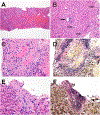
Figure 1.
Figure 1.
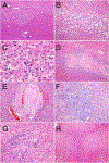
Case 1, biopsy. Panel A. The lesion shows patchy but striking sinusoidal dilatation and congestion. Panel B. At low power magnification, there were areas of subtle increase in the number of portal tracts. In this image taken with a 20X lens, 4 closely approximated portal tracts are evident (arrows). Panel C. A thrombosed central vein. Panel D. A VVG shows the same thrombosed vein (same field as prior image). Panel E. The central vein is inconspicuous and shows elastotic change. Panel F. A VVG stain suggests this could be a remotely thrombosed central vein.
Figure 2.
Figure 2.
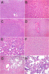
Case 2, resection. Panel A. The lesion (upper image) blends almost imperceptibly into the background liver. A thin line has been added to separate the lesion from the background liver. Panel B. At low power magnification, there is striking but patchy sinusoidal dilatation. Panel C. Intralobular arteries were present. Panel D. Multiple vessels in the portal tracts are thrombosed. In some portal tracts, the vessels were significantly distorted as well as occluded. Panel E. A nearly occluded portal vein at higher magnification. Panel F. Many portal tracts showed bile ductular proliferation. 1G. Prominent arteries were present in many portal tracts. 1H. The background liver also showed nodular regenerative hyperplasia.
Figure 3.
Figure 3.
Case 3, resection. Panel A. At low power, the lesion mimics an inflammatory hepatic adenoma. Panel B. At higher power magnification, however, the lesion has true portal tracts, instead of the faux portal tracts commonly present in inflammatory hepatic adenomas. Panel C. A remotely thrombosed portal vein is seen. Panel D. Many portal tracts have prominent, hyperplastic arteries.
Figure 4.
Figure 4.
Case 8, needle biopsy. Panel A. At low power, there is marked sinusoidal dilatation. Panel B. A thrombosed central vein is present. Panel C. The portal tracts showed patchy bile ductular proliferation. Panel D. One of the portal tracts shows a portal vein with an unusual morphology.
Figure 5.
Figure 5.
Follow-up resection on case 8. Panel A. At low power, the lesion (upper part of panel) blends imperceptibly with the background liver (lower part of image). Panel B. The lesion shows patchy, marked sinusoidal dilatation. Panel C. The portal tracts varied in their appearance throughout the lesion and were irregularly distributed. In this large field taken with a 2X lens, no portal tract were evident. Panel D. In this image, the portal vein is small, but the portal tract otherwise appears normal. Panel E. This portal tract has largely been resorbed, leaving only a remnant. Panel F. Other lesions in the background liver included three regenerative nodules with a solid appearance. Panel G. One pseudotumor lesion had at its periphery multiple foci of telangiectatic vessels. Panel F. A higher-magnification image of the telangiectatic vessels.

References

    1. Torbenson MS. Atlas of liver pathology : a pattern-based approach. Wolters Kluwer: Philadelphia, 2020. xii, 475 pages pp.
    1. Torbenson MS. Biopsy interpretation of the liver. Wolters Kluwer Health: Philadelphia, 2015. vii, 534 pages pp.
    1. Singhi AD, Maklouf HR, Mehrotra AK, et al. Segmental atrophy of the liver: a distinctive pseudotumor of the liver with variable histologic appearances. Am J Surg Pathol 2011;35:364–71. - PubMed
    1. Locke JE, Choti MA, Torbenson MS, Horton KM, Molmenti EP. Inflammatory pseudotumor of the liver. J Hepatobiliary Pancreat Surg 2005;12:314–6. - PubMed
    1. Taguchi K, Kuroda S, Kobayashi T, et al. Pseudolymphoma of the liver: a case report and literature review. Surg Case Rep 2015;1:107. - PMC - PubMed

Publication types