. 2020 Mar 30:9:e53387.
doi: 10.7554/eLife.53387.

A branched heterochronic pathway directs juvenile-to-adult transition through two LIN-29 isoforms

Affiliations

A branched heterochronic pathway directs juvenile-to-adult transition through two LIN-29 isoforms

Chiara Azzi et al. Elife. .

Abstract

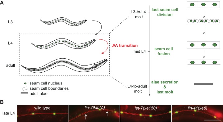
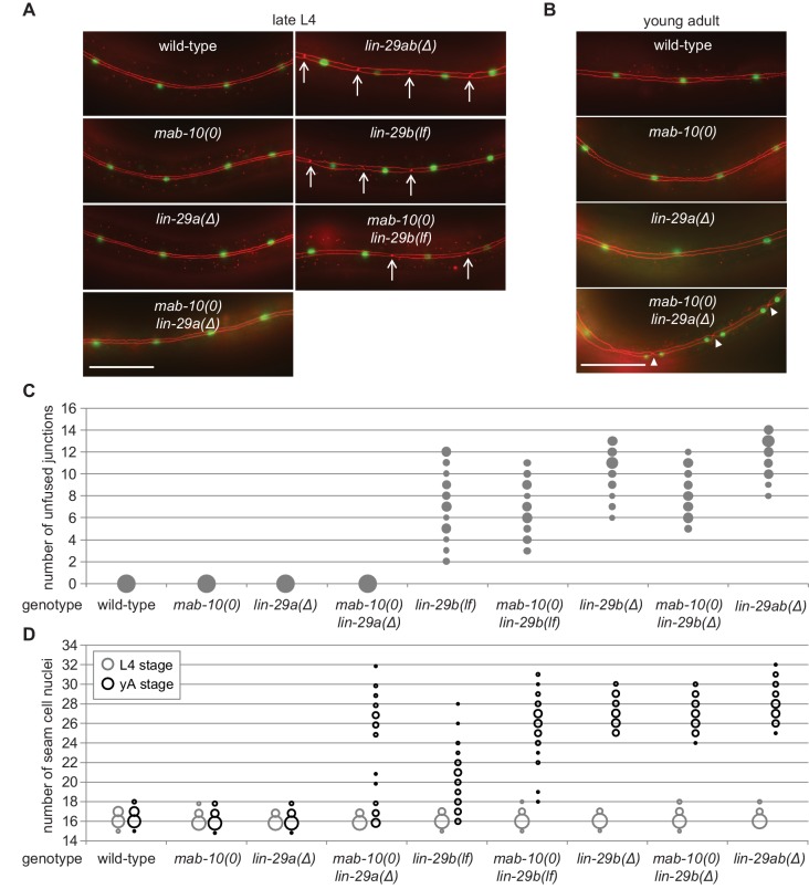
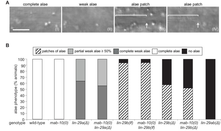
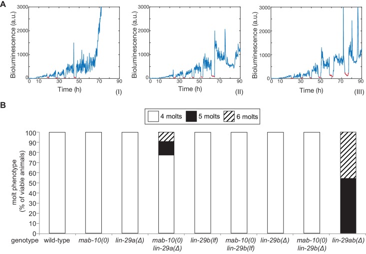
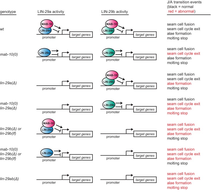
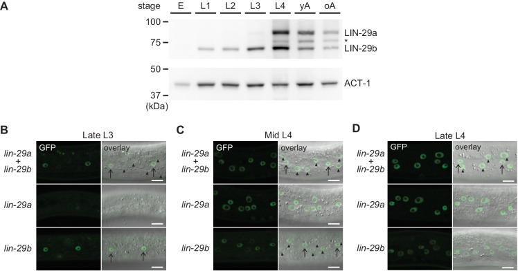
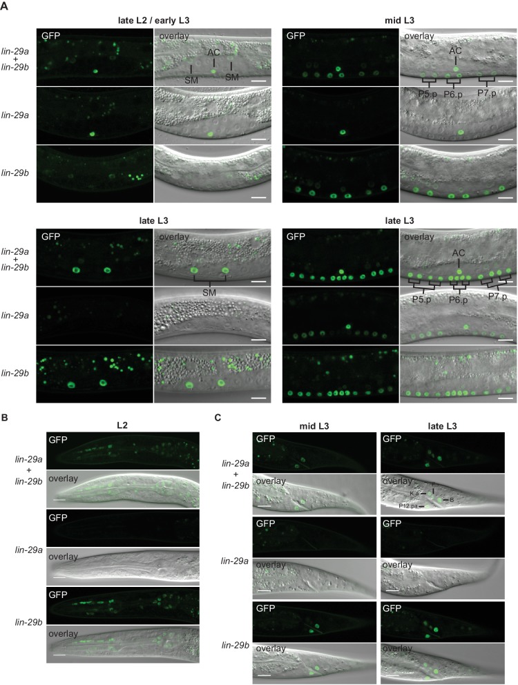
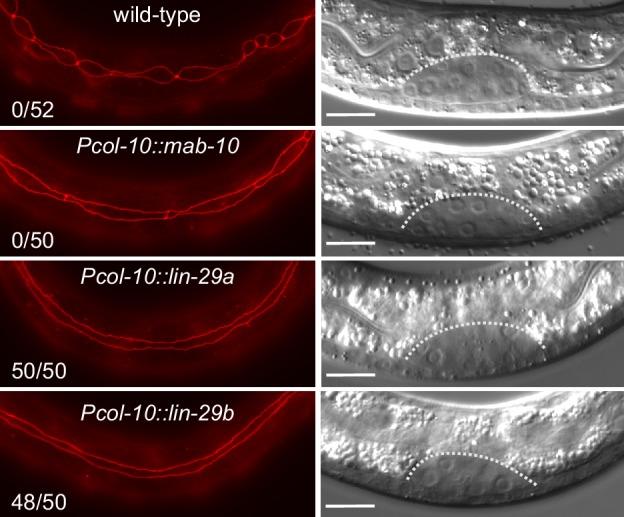
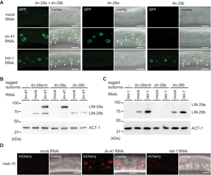
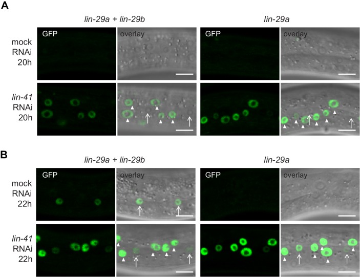
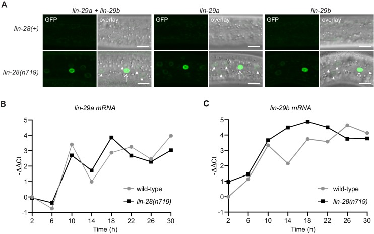
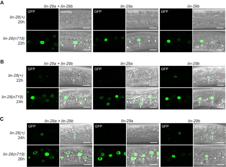
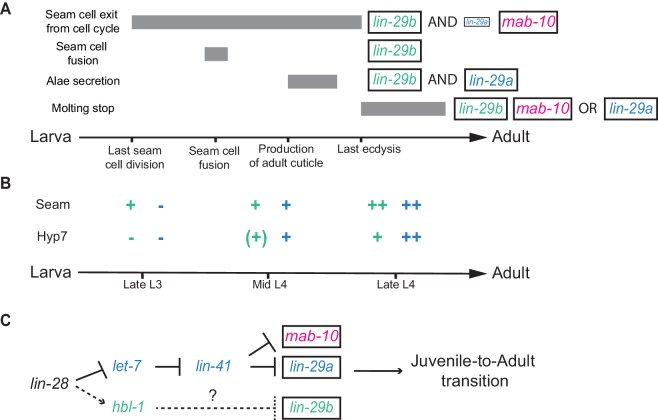
Robust organismal development relies on temporal coordination of disparate physiological processes. In Caenorhabditis elegans, the heterochronic pathway controls a timely juvenile-to-adult (J/A) transition. This regulatory cascade of conserved proteins and small RNAs culminates in accumulation of the transcription factor LIN-29, which triggers coordinated execution of transition events. We report that two LIN-29 isoforms fulfill distinct functions. Functional specialization is a consequence of distinct isoform expression patterns, not protein sequence, and we propose that distinct LIN-29 dose sensitivities of the individual J/A transition events help to ensure their temporal ordering. We demonstrate that unique isoform expression patterns are generated by the activities of LIN-41 for lin-29a, and of HBL-1 for lin-29b, whereas the RNA-binding protein LIN-28 coordinates LIN-29 isoform activity, in part by regulating both hbl-1 and lin-41. Our findings reveal that coordinated transition from juvenile to adult involves branching of a linear pathway to achieve timely control of multiple events.

Keywords: C. elegans; developmental biology; developmental timing; epidermis; heterochronic; molt; puberty; terminal differentiation.


Conflict of interest statement

CA, AN, HG No competing interests declared, FA The author is now affiliated with CSL Behring, Research, CSL Biologics Research Center, however all work was conducted when affiliated with Friedrich Miescher Institute for Biomedical Research and University of Basel.

Figures

Figure 1.
Figure 1.. Uncoupling of coordinated execution of J/A transition events in let-7 and lin-41 mutant animals.
(A) Schematic representation of juvenile-to-adult (J/A) transition events in the C. elegans epidermis: final division of seam cells (square-shaped cells with green nuclei) at the L3-to-L4 molt; seam cell fusion into a syncytium during mid-L4 stage; synthesis of an adult cuticle containing lateral alae (three horizontal bars) at the L4-to-adult molt; and a subsequent exit from the molting cycle. (B) Micrographs of late L4-stage animals of indicated genotypes expressing scm::gfp (green, marking seam cells) and ajm-1::mCherry (red, marking hypodermal cell junctions). Arrows indicate cell boundaries between unfused cells. Representative of n > 20. Scale bar: 50 μm.
Figure 2.
Figure 2.. Generation of lin-29a and lin-29b isoform-specific mutants.
(A) Schematic representation of the lin-29 and mab-10 genomic regions. Mutant alleles and endogenously tagged alleles used in this study are indicated. Insertion of agfp::3xflag-encoding sequence at the 5' end specifically tags LIN-29a at its N-terminus, while insertion at the 3’ end tags both isoforms at the shared C-terminus. Insertion of this C-terminal tag in a lin-29(xe40[lin-29a(Δ)]) genetic background yields specific tagging of LIN-29b. Allele numbers refer to modifications in otherwise wild-type backgrounds; numbers for equivalent mutations in the endogenously tagged backgrounds, used for protein detection by microscopy and Western blotting, are provided in the Key resources table. (B) Western blot of C-terminally GFP::3xFLAG-tagged endogenous LIN-29a and LIN-29b proteins in the different mutant backgrounds using anti-FLAG antibody. Animals were grown for 36 hr at 25°C to the late L4 stage. Actin-1 is used as loading control.
Figure 3.
Figure 3.. LIN-29b has a fundamental role in the regulation of early J/A transition events.
(A–B) Micrographs of late L4 stage (A) and young adult (B) animals of indicated genotype expressing scm::gfp (green, marking seam cells) and ajm-1::mCherry (red, marking seam cell boundaries). Arrows indicate cell boundaries between unfused seam cells, arrowheads indicate newly formed seam cell boundaries. Scale bars: 50 μm. (C) Quantification of unfused seam cell junctions inL4 larval stage animals of the indicated genetic backgrounds. Areas of bubbles represent the percentage of worms with the respective number of unfused junctions (n > 20 for each genotype). (D) Quantification of seam cell numbers in L4 larval stage and young adult (yA) animals of the indicated genetic backgrounds. Areas of bubbles represent the percentage of worms with the respective number of seam cells (n = 20 for L4, n > 50 for yA worms per genotype).
Figure 4.
Figure 4.. LIN-29a and LIN-29b, but not MAB-10, are required for wild-type alae formation.
(A) Micrographs illustrating categories of alae structures observed on the cuticle of wild-type (I) and mutant (II – IV) young adult animals. The example pictures show (I) wild-type N2, (II) mab-10(0) lin-29a(Δ), (III) mab-10(0) lin-29b(lf) and (IV) mab-10(0) lin-29b(Δ) animals, respectively. Scale bar: 10 μm. (B) Quantification of different alae structures in young adult worms of indicated genotypes (n > 30).
Figure 5.
Figure 5.. The molting cycle is regulated by both LIN-29 isoforms.
(A) Examples of luciferase assay traces revealing four (I), five (II) or six (III) molts through a drop in luciferase signal (red segment). Examples are from wild-type (I) and lin-29ab(Δ) (II-III). (B) Quantification of the number of molts in animals of the indicated genotypes (n > 20) based on the assay shown in (A). A fraction of mab-10(0) lin-29a(∆) lin-29b(∆) and mab-10(0) lin-29b(∆) animals die at the J/A transition ; these animals were censored and not included in the quantification.
Figure 6.
Figure 6.. The LIN-29a-specific domain is dispensable for the execution of the J/A transition.
(A) Western blot of endogenous C-terminally GFP::3xFLAG-tagged LIN-29a and LIN-29b proteins in the lin-29a(ΔN) background (HW2408) using an anti-FLAG antibody. Actin-1 is used as loading control. (B) Seam cell number quantification in L4 larval stage and young adult (yA) animals of the indicated genetic backgrounds (n > 25 for L4, n > 25 for yA worms per genotype). The data for lin-29a(Δ) and mab-10(0) lin-29a(Δ) is re-plotted from Figure 2 for comparison. (C) Quantification of different alae structures in young adult worms of indicated genotypes (n > 20). The data for lin-29a(Δ) and mab-10(0) lin-29a(Δ) is re-plotted from Figure 3 for comparison. (D) Quantification of the number of molts for animals of indicated genotypes (n > 20). The data for lin-29a(Δ) and mab-10(0) lin-29a(Δ) is re-plotted from Figure 3 for comparison. In (B – D), lin-29(∆N) is lin-29(xe200).
Figure 6—figure supplement 1.
Figure 6—figure supplement 1.. Summary of the J/A transition phenotypes seen for different permutations of lin-29a, lin-29b, and mab-10 mutations.
Note that extra seam cell divisions in mab-10(0) mutant animals occur only in older adults. Some older mab-10 mutant adults may also undergo extra molts (Harris and Horvitz, 2011), although we did not observe this.
Figure 6—figure supplement 2.
Figure 6—figure supplement 2.. Characterization of lin-29a(∆N) expression and function.
(A) Schematic representation of the lin-29a(ΔN) deletion. (B) Micrographs of late L4-stage animals of indicated genotypes expressing scm::gfp (green, marking seam cells) and ajm-1::mCherry (red, marking hypodermal cell junctions). In both lin-29a(xe200[lin-29a(ΔN)]) and mab-10(0) lin-29a(xe200) animals, fusion occurs normally. Scale bar: 50 μm. (C) Western blot of C-terminally GFP::3xFLAG-tagged endogenous LIN-29a and LIN-29b proteins in a wild-type and the lin-29a(ΔN) background (HW2408), respectively, using anti-FLAG antibody. Animals were grown for 20 hr at 25°C to the L3 stage on mock RNAi and lin-41 RNAi bacteria, respectively. Both LIN-29a and LIN-29a(∆N) accumulate upon knock-down of lin-41. Actin-1 is used as a loading control. (D) Confocal images of endogenously tagged LIN-29 protein isoforms in the wild type and lin-29a(ΔN) background (HW2408) in the epidermis of animals at the indicated developmental stages. Animals were staged by examination of gonad development. Arrows indicate seam cell, arrowheads hyp7 nuclei. Scale bars: 10 μm.
Figure 7.
Figure 7.. Spatial and temporal expression pattern of LIN-29a and LIN-29b.
(A) Western blot of C-terminally GFP::3xFLAG-tagged endogenous LIN-29a and LIN-29b proteins in different developmental stages using an anti-FLAG antibody. Actin-1 is used as a loading control. The asterisk indicates a band of unclear origin. (B–D) Confocal images of endogenously tagged LIN-29 isoforms in the epidermis of animals at the indicated developmental stages, which were confirmed by examination of gonad development. Arrows indicate seam cell, arrowheads hyp7 nuclei. Scale bars: 10 μm.
Figure 7—figure supplement 1.
Figure 7—figure supplement 1.. Expression of lin-29 isoforms.
(A) Confocal images of endogenously tagged LIN-29 protein isoforms in the region of the vulva and the uterus at the indicated developmental stages. At the L2-to-L3 molt (A), lin-29a is expressed in the anchor cell (AC), while lin-29b is weakly expressed in the sex myoblasts (SMs). In mid-L3 stage worms, the six daughters of the VPCs P5.p-P7.p express lin-29b. At the late L3 stage, lin-29b is strongly expressed in the sex myoblast (SM) daughters and all 12 granddaughters of the VPCs P5.p-P7.p, while LIN-29a specifically accumulates in the granddaughters of P5.p and P7.p, but not in those of P6.p. Scale bars: 10 μm. (B) Confocal images of endogenously tagged LIN-29 protein isoforms in the pharynx of L2 stage animals at the indicated developmental stages. lin-29b is expressed in the pharynx throughout larval and adult development. Scale bars: 10 μm. (C) Confocal images of endogenously tagged LIN-29 protein isoforms in the tail region at the indicated developmental stages. LIN-29b first accumulates in the two rectal cells B and P12.pa, before accumulating in the four additional rectal cells F, K.a, K’ and U (the latter two are not visible in this focal plane). LIN-29a is not detected in these cells. Scale bars: 10 μm.
Figure 8.
Figure 8.. Precocious LIN-29 accumulation is sufficient for seam cell fusion in early L2 worms.
Micrographs of early L2 animals expressing ajm-1::mCherry (red, marking hypodermal cell junctions) and the indicated transgenes (‘wild-type’: no transgene). Animal stage was confirmed by gonad morphology as shown in the DIC pictures. Numbers indicate fractions of animals with complete precocious seam cells fusion. Scale bars: 10 μm.
Figure 9.
Figure 9.. lin-29a and lin-29b expression are specifically regulated by LIN41 and HBL-1, respectively.
(A) Confocal images of endogenously tagged LIN-29 protein isoforms in the epidermis (strains HW1822, HW1826, HW1835). Animals were grown at 25°C for 20 hr to the early L3 stage on RNAi bacteria as indicated. Arrows indicate seam cell, arrowheads hyp7 nuclei. Scale bars: 10 μm. (B–C) Western blot of GFP::3xFLAG-tagged endogenous LIN-29a and LIN-29b proteins using an anti-FLAG antibody. Actin-1 is used as a loading control. Animals were grown for 20 hr at 25°C to the early L3 stage on RNAi bacteria as indicated. (D) Confocal images of endogenously tagged MAB-10 protein in the epidermis. Animals were grown at 25°C for 20 hr on lin-41, hbl-1 or mock RNAi bacteria. Arrowheads indicate hyp7 cells, arrows seam cells. Scale bars: 10 μm.
Figure 9—figure supplement 1.
Figure 9—figure supplement 1.. Expression of lin-29a on lin-41 RNAi over time.
(A–B) Confocal images of endogenously tagged LIN-29 protein isoforms in the epidermis (strains HW1822, HW1826, HW1835). Animals were grown at 25°C for 20 hr (A) or 22 hr (B) to the early L3 stage on RNAi bacteria as indicated. Arrows indicate seam cell, arrowheads hyp7 nuclei. Scale bars: 10 μm.
Figure 9—figure supplement 2.
Figure 9—figure supplement 2.. mRNA levels of lin-29b in worms grown on lin-41 and hbl-1 RNAi over time.
RT-qPCR analysis to measure the -∆∆Ct of lin-29b mRNA levels (normalized by act-1 mRNA levels) over time. Wild-type animals exposed to lin-41 and hbl-1 RNAi compared to mock RNAi.
Figure 10.
Figure 10.. LIN-28 coordinates both lin-29a and lin-29b regulation.
(A) Confocal images of endogenously tagged LIN-29 protein isoforms in wild-type or lin-28(n719) background in the epidermis (strains HW1822, HW1826, HW1835, HW1924, HW1925, HW1926). Animals were grown at 25°C for 20 hr (control strains) and 22 hr (lin-28(n719) strains) to reach an equivalent early L3 developmental stage. Arrows indicate seam cell, arrowheads hyp7 nuclei. Scale bars: 10 μm. (B) RT-qPCR analysis to measure the -∆∆Ct of lin-29a mRNA levels in wild-type and lin-28(n719) mutant background (normalized by act-1 mRNA levels) over time. (C) RT-qPCR analysis to measure the -∆∆Ct of lin-29b mRNA levels in wild-type and lin-28(n719) mutant background (normalized by act-1 mRNA levels) over time.
Figure 10—figure supplement 1.
Figure 10—figure supplement 1.. Expression of lin-29a and lin-29b in wild-type and lin-28 mutant animals over time.
(A–C) Confocal images of endogenously tagged LIN-29 protein isoforms in wild-type (lin-28(+)) or lin-28(n719) background in the epidermis (strains HW1822, HW1826, HW1835, HW1924, HW1925, HW1926). Animals were grown at 25°C for 20 hr, 22 hr and 24 hr (A, B, and C, resp., for control strains) and 22 hr, 24 hr and 26 hr (A, B, and C, resp., for lin-28(n719) strains) to reach an equivalent developmental stage. Arrows indicate seam cell, arrowheads hyp7 nuclei. Scale bars: 10 μm.
Figure 11.
Figure 11.. Summary.
(A) Illustration of the contributions of LIN-29 isoforms and the co-factor MAB-10 to the different J/A transition events. The arrow represents developmental time with relevant events indicated. Filled boxes indicate the duration of a specific J/A transition event. The exact time point when seam cells exit the cell cycle is unknown and might occur at any time after the last division at the L3/L4 molt and before the L4/adult molt. Although adults do not molt, it is unknown when exit from the molting cycle occurs. (B) Schematic depiction of lin-29a (blue) and lin-29b (green) expression patterns in seam cells and hyp7. Relevant developmental stages are indicated on the arrow representing developmental time. (C) Revised model of the heterochronic pathway. Two parallel arms of the heterochronic pathway exert their functions through distinct LIN-29 isoforms. Their activities are coordinated throught the upstream function of LIN-28. Dashed arrow indicate indirect regulation which involve let-7 sisters miRNAs in hyp7 (Abbott et al., 2005) and LIN-46 in the seam cells (Ilbay and Ambros, 2019; Ilbay et al., 2019).
Author response image 1.
Author response image 1.. Replicate Western blot experiments.
Author response image 2.
Author response image 2.. Vulval phenotypes upon precocious lin-29 expression.
Author response image 3.
Author response image 3.. Seam cell fusion upon precocious lin-29 expression in only the seam.

References

    1. Abbott AL, Alvarez-Saavedra E, Miska EA, Lau NC, Bartel DP, Horvitz HR, Ambros V. The let-7 MicroRNA family members mir-48, mir-84, and mir-241 function together to regulate developmental timing in Caenorhabditis elegans. Developmental Cell. 2005;9:403–414. doi: 10.1016/j.devcel.2005.07.009. - DOI - PMC - PubMed
    1. Abete-Luzi P, Fukushige T, Yun S, Krause MW, Eisenmann DM. New roles for the heterochronic transcription factor LIN-29 in cuticle maintenance and lipid metabolism at the Larval-to-Adult transition in Caenorhabditis elegans. Genetics. 2020;214:669–690. doi: 10.1534/genetics.119.302860. - DOI - PMC - PubMed
    1. Abete-Luzi P, Eisenmann DM. Regulation of C. elegans L4 cuticle collagen genes by the heterochronic protein LIN-29. Genesis. 2018;56:23106. doi: 10.1002/dvg.23106. - DOI - PMC - PubMed
    1. Abrahante JE, Daul AL, Li M, Volk ML, Tennessen JM, Miller EA, Rougvie AE. The Caenorhabditis elegans hunchback-like gene lin-57/hbl-1 controls developmental time and is regulated by microRNAs. Developmental Cell. 2003;4:625–637. doi: 10.1016/S1534-5807(03)00127-8. - DOI - PubMed
    1. Abreu AP, Dauber A, Macedo DB, Noel SD, Brito VN, Gill JC, Cukier P, Thompson IR, Navarro VM, Gagliardi PC, Rodrigues T, Kochi C, Longui CA, Beckers D, de Zegher F, Montenegro LR, Mendonca BB, Carroll RS, Hirschhorn JN, Latronico AC, Kaiser UB. Central precocious puberty caused by mutations in the imprinted gene MKRN3. New England Journal of Medicine. 2013;368:2467–2475. doi: 10.1056/NEJMoa1302160. - DOI - PMC - PubMed

Publication types

MeSH terms