. 2020 Apr 24;23(4):100987.
doi: 10.1016/j.isci.2020.100987. Epub 2020 Mar 16.

Surface LSP-1 Is a Phenotypic Marker Distinguishing Human Classical versus Monocyte-Derived Dendritic Cells

Affiliations

Surface LSP-1 Is a Phenotypic Marker Distinguishing Human Classical versus Monocyte-Derived Dendritic Cells

Sandrine Moutel et al. iScience. .

Abstract

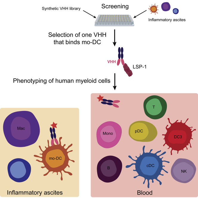
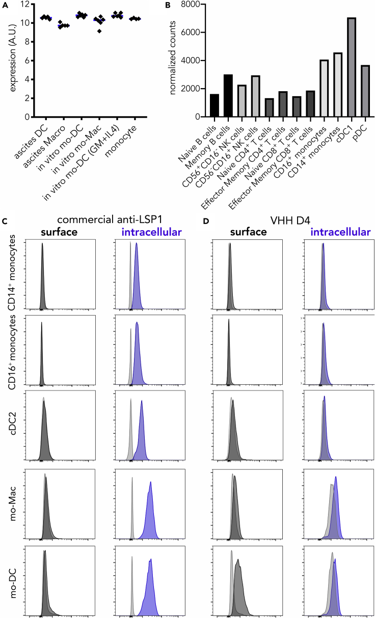
Human mononuclear phagocytes comprise several subsets of dendritic cells (DCs), monocytes, and macrophages. Distinguishing one population from another is challenging, especially in inflamed tissues, owing to the promiscuous expression of phenotypic markers. Using a synthetic library of humanized llama single domain antibodies, we identified a novel surface marker for human naturally occurring monocyte-derived DCs. Our antibody targets an extra-cellular domain of LSP-1, specifically on monocyte-derived DCs, but not on other leukocytes, in particular monocytes, macrophages, classical DCs, or the recently described blood DC3 population. Our findings will pave the way for a better characterization of human mononuclear phagocytes in pathological settings.

Keywords: Molecular Biology.


Conflict of interest statement

Declaration of Interests S.M., S.A., F.P., and E.S. are co-inventors of a patent entitled "New anti-LSP1 antibody" (PCT/EP2017/0761). S.M. and F.P. are co-inventors of a patent that covers the commercial use of the library (WO/2015/063331). The authors declare no other competing interest. The authors adhere to Cell Press policy on sharing materials.

Figures

None
Graphical abstract
Figure 1
Figure 1
Identification of a VHH Specific for Monocyte-Derived Dendritic Cells (A) Strategy for identifying novel surface markers for ascites dendritic cells (DCs). (B) Ascites cells were stained with VHH D4. Stainings for DC and macrophages (Macro) are shown (representative of five individual donors). Gray shaded histograms are fluorescence-minus-one controls. (C) Monocytes were cultured with M-CSF, IL-4, and TNFa to generate DC (mo-DC) and macrophages (mo-Mac) or with GM-CSF and IL-4 to generate DC (GM + IL4 mo-DC). Cells were stained with VHH D4. Gray shaded histograms are fluorescence-minus-one controls. Representative of four individual donors.
Figure 2
Figure 2
VHH D4 Does Not Stain Classical Dendritic Cell Populations (A) Tonsil DC and macrophages were stained with VHH D4. Gray shaded histograms are fluorescence-minus-one controls. Representative of two individual donors. Peripheral blood cells (B and C) or ascites cells (C) were stained with VHH D4. Gray shaded histograms are fluorescence-minus-one controls. Representative of five (blood) or four (ascites) individual donors. (C) Gating strategy for analyzing HLA-DR+ cells, including CD14+ monocytes, cDC2, and DC3, is shown. Staining of ascites DC is shown as a positive control.
Figure 3
Figure 3
Identification of LSP-1 as the Target of VHH D4 In vitro-generated mo-DC or mo-Mac were incubated with VHH D4, then lysed. Immuno-precipitation was performed on cell lysate using the streptavidin-binding peptide tag. MW, molecular weight. Immuno-precipitated material was analyzed by western blot. Staining was performed using streptavidin-HRP (A) or a commercial anti-LSP-1 antibody (B).
Figure 4
Figure 4
VHH D4 Recognizes a Surface Epitope of LSP-1 Specifically Expressed by mo-DC (A) Expression levels (arbitrary units) of LSP1 in ascites DC; ascites macrophages; in vitro mo-DC and mo-Mac generated with M-CSF, IL-4, and TNFα; in vitro mo-DC generated with GM-CSF and IL-4; and blood CD14+ monocytes. Each dot represents an individual donor. Median is shown. Affymetrix data from dataset GSE102046. (B) Normalized counts of LSP1 expression in selected human blood cells. RNA-seq data from the Human Cell Atlas (http://immunecellatlas.net). (C and D) Monocytes were cultured with M-CSF, IL-4, and TNFα to generate DC (mo-DC) and macrophages (mo-Mac). Peripheral blood myeloid cells were also analyzed (monocytes and cDC2). Cells were stained with a commercial anti-LSP-1 antibody (C) or VHH D4 (D), with membrane permeabilization (intracellular) or not (surface). Gray shaded histograms are fluorescence-minus-one controls. Representative of five (blood) or six (in vitro-generated) individual donors.

References

    1. Alcantara-Hernandez M., Leylek R., Wagar L.E., Engleman E.G., Keler T., Marinkovich M.P., Davis M.M., Nolan G.P., Idoyaga J. High-dimensional phenotypic mapping of human dendritic cells reveals interindividual variation and tissue specialization. Immunity. 2017;47:1037–1050.e6. - PMC - PubMed
    1. Bakdash G., Buschow S.I., Gorris M.A., Halilovic A., Hato S.V., Skold A.E., Schreibelt G., Sittig S.P., Torensma R., Duiveman-De Boer T. Expansion of A Bdca1+Cd14+ myeloid cell population in melanoma patients may attenuate the efficacy of dendritic cell vaccines. Cancer Res. 2016;76:4332–4346. - PubMed
    1. Coillard A., Segura E. In vivo differentiation of human monocytes. Front. Immunol. 2019;10:1907. - PMC - PubMed
    1. Cytlak U., Resteu A., Pagan S., Green K., Milne P., Maisuria S., Mcdonald D., Hulme G., Filby A., Carpenter B. SSRN; 2019. Differential Irf8 Requirement Defines Two Pathways of Dendritic Cell Development in Humans. - PMC - PubMed
    1. Durand M., Walter T., Pirnay T., Naessens T., Gueguen P., Goudot C., Lameiras S., Chang Q., Talaei N., Ornatsky O. Human lymphoid organ Cdc2 and macrophages play complementary roles in T follicular helper responses. J. Exp. Med. 2019;216:1561–1581. - PMC - PubMed