. 2020 Jun:36:100966.
doi: 10.1016/j.molmet.2020.02.009. Epub 2020 Feb 18.

Wild-type IDH2 contributes to Epstein-Barr virus-dependent metabolic alterations and tumorigenesis

Affiliations

Wild-type IDH2 contributes to Epstein-Barr virus-dependent metabolic alterations and tumorigenesis

Feng Shi et al. Mol Metab. 2020 Jun.

Abstract

Objective: Epstein-Barr virus (EBV) is a well-recognized oncogenic virus that can induce host cell metabolic reprogramming and tumorigenesis by targeting vital metabolic enzymes or regulators. This study aims to explore the role of wild-type isocitrate dehydrogenase 2 (IDH2) in metabolic reprogramming and tumorigenesis induced by EBV-encoded latent membrane protein 1 (LMP1).

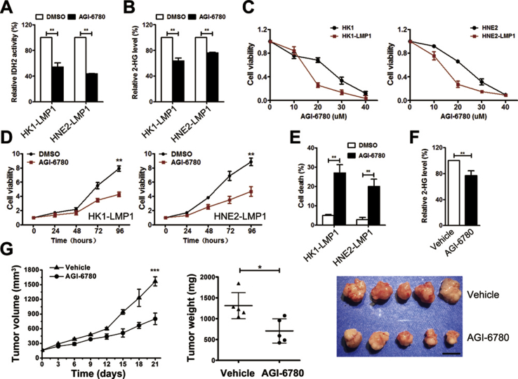
Methods: Mechanistic dissection of wild-type IDH2 in EBV-LMP1-induced tumorigenesis was investigated using western blotting, real-time polymerase chain reaction (PCR), immunochemistry, chromatin immunoprecipitation (ChIP), and luciferase assay. The role of wild-type IDH2 was examined by cell viability assays/Sytox Green staining in vitro and xenograft assays in vivo.

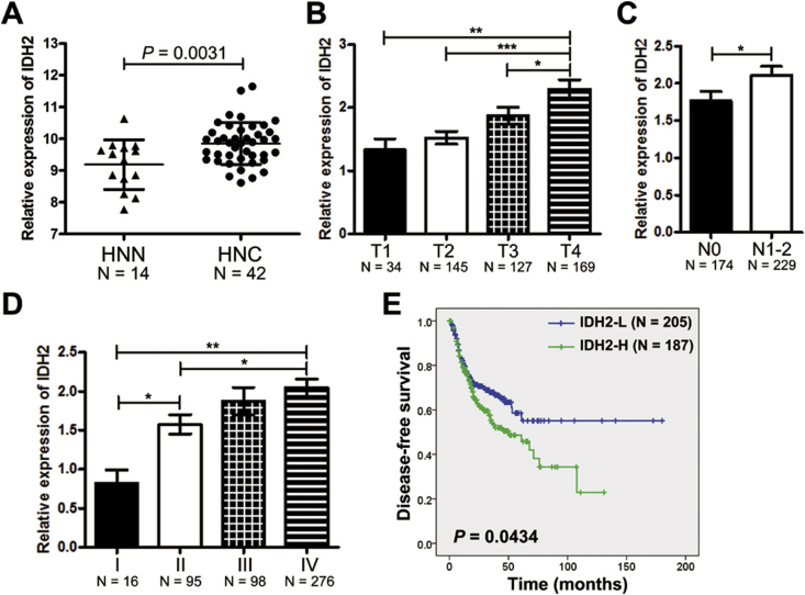
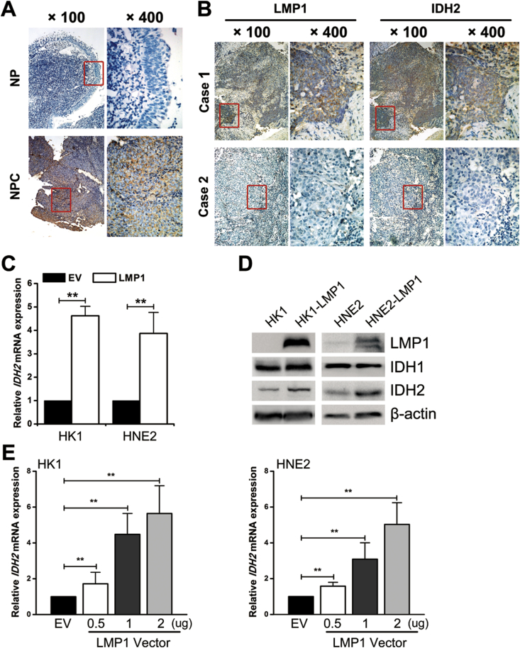
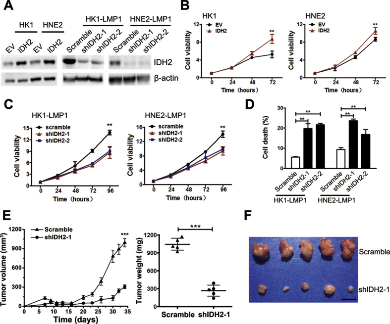
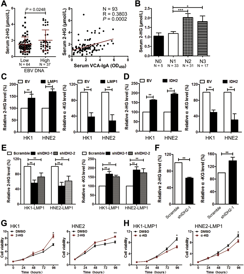
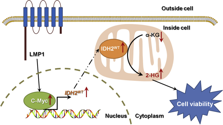
Results: IDH2 over-expression is a prognostic indicator of poorer disease-free survival for patients with head and neck squamous cell carcinoma (HNSCC). IDH2 expression is also upregulated in nasopharyngeal carcinoma (NPC, a subtype of HNSCC) tissues, which is positively correlated with EBV-LMP1 expression. EBV-LMP1 contributes to NPC cell viability and xenograft tumor growth mediated through wild-type IDH2. IDH2-dependent changes in intracellular α-ketoglutarate (α-KG) and 2-hydroxyglutarate (2-HG) contribute to EBV-LMP1-induced tumorigenesis in vitro and in vivo. Elevated serum 2-HG level is associated with high EBV DNA and viral capsid antigen-immunoglobulin A (VCA-IgA) levels in patients with NPC. A significantly positive correlation exists between serum 2-HG level and regional lymph node metastases of NPC. EBV-LMP1 enhances the binding of c-Myc with the IDH2 promoter and transcriptionally activates wild-type IDH2 through c-Myc. Targeting IDH2 decreased intracellular 2-HG levels and survival of EBV-LMP1-positive tumor cells in vitro and in vivo.

Conclusions: Our results demonstrate that the EBV-LMP1/c-Myc/IDH2WT signaling axis is critical for EBV-dependent metabolic changes and tumorigenesis, which may provide new insights into EBV-related cancer diagnosis and therapy.

Keywords: Epstein-Barr virus; Isocitrate dehydrogenase 2; Metabolic reprogramming; Nasopharyngeal carcinoma; Tumorigenesis.


Figures

Image 1
Graphical abstract
Figure 1
Figure 1
IDH2 over-expression in HNSCC. A, IDH2 expression in HNSCC tissues (HNC, N = 42) and normal tissues (N = 14) from the online GENT database. IDH2 expression was compared among different stages of B, pathologic T stage, C, regional lymph node metastases N stage, and D, AJCC clinical stage in patients with HNSCC from the TCGA database. E, Kaplan–Meier survival curves of patients with HNSCC with IDH2 expression from the TCGA database. Data are presented as means ± S.D.; ∗, p < 0.05; ∗∗, p < 0.01; ∗∗∗, p < 0.001.
Figure 2
Figure 2
EBV-LMP1 promotes wild-type IDH2 overexpression in NPC. A, representative IHC photos for the expression of IDH2 in nasopharyngitis (NP) and nasopharyngeal carcinoma (NPC) tissues. B, representative IHC photos for the expression of LMP1 and IDH2 in consecutive sections of NPC tissues. C, IDH2 mRNA expression was determined by RT-PCR (columns = mean; bars = S.D.; N = 3; ∗∗, p < 0.01). D, LMP1, IDH1, IDH2 protein expression was detected by western blot analysis, and β-actin served as a loading control. E, HK1 (left) and HNE2 (right) cells were transiently transfected with 0.5, 1, and 2 μg of LMP1 vector. IDH2 mRNA expression was determined by RT-PCR (columns = mean; bars = S.D.; N = 3; ∗∗, p < 0.01).
Figure 3
Figure 3
Wild-type IDH2 plays a key role in EBV-LMP1-induced tumorigenesis. A, IDH2 protein expression was detected by western blot analysis. EV, empty vector. B, cell viability was measured in empty vector- or IDH2-overexpressing cells using the MTS assay. C, viability was measured in cells transfected with scramble or IDH2 shRNA using the MTS assay. D, cell death was measured by Sytox Green staining in groups as indicated. E, tumor volume and weight were measured in xenografts in mice inoculated with HNE2-LMP1 cells transfected with scramble or IDH2 shRNA-1 (N = 5). F, photographs of xenograft tumors are shown (scale bar = 1 cm). Data are presented as means ± S.D.; ∗∗, p < 0.01; ∗∗∗, p < 0.001.
Figure 4
Figure 4
Overexpression of wild-type IDH2 is responsible for EBV-LMP1-modulated metabolic changes. A, the patients were divided into two groups based on their serum EBV DNA copy number: 1) low-level group = EBV DNA ≥1 × 103 copies and 2) high-level group = EBV DNA<1 × 103 copies. Serum 2-HG levels were compared between the two groups (left). The correlation between serum 2-HG levels and VCA-IgA levels was analyzed (right). B, serum 2-HG levels were compared among different stages of regional lymph node metastases N stage. The levels of 2-HG (left) and α-KG (right) were measured in C, NPC cells transfected with empty or LMP1 expressing vector, D, in NPC cells transfected with empty or IDH2 expressing vector, in E, NPC cells transfected with scramble or IDH2 shRNAs, and in F, xenograft tissues from mice inoculated with HNE2-LMP1 cells transfected with scramble or IDH2 shRNA-1. Viability was measured by MTS in cells treated with G, DMSO/1 mM octyl-2-HG, and H, cells treated with DMSO/1 mM octyl-α-KG. Data are presented as means ± S.D.; ∗, p < 0.05; ∗∗, p < 0.01; ∗∗∗, p < 0.001.
Figure 5
Figure 5
EBV-LMP1 facilitates IDH2 transcription through c-Myc. A, Myc-TA-luc and pRL-TK plasmids were co-transfected into NPC cells. The transcriptional activity of c-Myc was determined by the dual luciferase reporter assay. B, c-Myc and IDH2 protein expression was detected by western blot analysis in cells transfected with control or c-Myc siRNAs. C, IDH2 mRNA expression was measured by RT-PCR. D, ChIP analysis was used to detect the presence of c-Myc in the promoter region of IDH2 by using two primer sites. E, pGL3-IDH2 and pRL-TK plasmids were co-transfected into NPC cells. IDH2 promoter activity was determined by the dual luciferase reporter assay. F, IDH2 promoter activity was determined in cells transfected with control or c-Myc siRNAs. G, luciferase activity was determined in cells transfected with pGL3-Basic, pGL3-IDH2, and pGL3-IDH2-mut (deletion of the c-Myc-binding site from −1446 to −1437 bp in pGL3-IDH2). H, viability was measured in cells transfected with control or c-Myc siRNAs by using MTS. The levels of I, 2-HG and J, α-KG (J) were measured in NPC cells transfected with control or c-Myc siRNAs. Data are presented as means ± S.D.; ∗, p < 0.05; ∗∗, p < 0.01.
Figure 6
Figure 6
Targeting wild-type IDH2 inhibits cell viability and tumor growth. A, IDH activity and B, 2-HG levels were examined in NPC cells treated with DMSO/20 μM AGI-6780 for 24 h. C, viability was measured by MTS in cells treated with different concentrations of AGI-6780 (0–40 μM) for 48 h. D, viability was measured in cells treated with DMSO/10 μM AGI-6780. E, cell death was measured by Sytox Green staining in cells treated with DMSO/10 μM AGI-6780 for 48 h. F, levels of 2-HG were measured in xenograft tissues from mice inoculated with HNE2-LMP1 cells treated with corn oil (vehicle)/AGI-6780 (150 mg/kg). G, tumor volume (left) and tumor weight (middle) were measured in xenografts from mice inoculated with HNE2-LMP1 cells treated with vehicle/AGI-6780 (N = 5). Photographs of xenograft tumors are shown (right, scale bar = 1 cm). Data are presented as means ± S.D.; ∗, p < 0.05; ∗∗, p < 0.01; ∗∗∗, p < 0.001.
Figure 7
Figure 7
The EBV-LMP1/c-Myc/IDH2WT signaling axis is critical for EBV-LMP1–dependent metabolic changes and tumorigenesis.

References

    1. Plummer M., de Martel C., Vignat J., Ferlay J., Bray F., Franceschi S. Global burden of cancers attributable to infections in 2012: a synthetic analysis. Lancet Global Health. 2016;4(9):e609–e616. - PubMed
    1. Khan G., Hashim M.J. Global burden of deaths from Epstein-Barr virus attributable malignancies 1990-2010. Infectious Agents and Cancer. 2014;9(1):38. - PMC - PubMed
    1. Cao Y. EBV based cancer prevention and therapy in nasopharyngeal carcinoma. NPJ Precision Oncology. 2017;1(1):10. - PMC - PubMed
    1. Tsao S.W., Tsang C.M., To K.F., Lo K.W. The role of Epstein-Barr virus in epithelial malignancies. The Journal of Pathology. 2015;235(2):323–333. - PMC - PubMed
    1. Yang H., Ye D., Guan K.L., Xiong Y. IDH1 and IDH2 mutations in tumorigenesis: mechanistic insights and clinical perspectives. Clinical Cancer Research. 2012;18(20):5562–5571. - PMC - PubMed

Publication types

MeSH terms

Substances