Review
. 2020 Mar 2;13(2):dmm043638.
doi: 10.1242/dmm.043638.

Biomarkers for Duchenne muscular dystrophy: myonecrosis, inflammation and oxidative stress

Affiliations
Review

Biomarkers for Duchenne muscular dystrophy: myonecrosis, inflammation and oxidative stress

Miranda D Grounds et al. Dis Model Mech. .

Abstract

Duchenne muscular dystrophy (DMD) is a lethal, X-linked disease that causes severe loss of muscle mass and function in young children. Promising therapies for DMD are being developed, but the long lead times required when using clinical outcome measures are hindering progress. This progress would be facilitated by robust molecular biomarkers in biofluids, such as blood and urine, which could be used to monitor disease progression and severity, as well as to determine optimal drug dosing before a full clinical trial. Many candidate DMD biomarkers have been identified, but there have been few follow-up studies to validate them. This Review describes the promising biomarkers for dystrophic muscle that have been identified in muscle, mainly using animal models. We strongly focus on myonecrosis and the associated inflammation and oxidative stress in DMD muscle, as the lack of dystrophin causes repeated bouts of myonecrosis, which are the key events that initiate the resultant severe dystropathology. We discuss the early events of intrinsic myonecrosis, along with early regeneration in the context of histological and other measures that are used to quantify its incidence. Molecular biomarkers linked to the closely associated events of inflammation and oxidative damage are discussed, with a focus on research related to protein thiol oxidation and to neutrophils. We summarise data linked to myonecrosis in muscle, blood and urine of dystrophic animal species, and discuss the challenge of translating such biomarkers to the clinic for DMD patients, especially to enhance the success of clinical trials.

Keywords: Biomarkers; Blood; DMD; Dogs; Dystrophic mice; Inflammation; Muscle necrosis; Neutrophils; Oxidative stress; Rats; Urine.


Conflict of interest statement

Competing interestsThe authors declare no competing or financial interests.

Figures

Fig. 1.
Fig. 1.
Necrosis of dystrophic skeletal muscle and associated cellular events. (A) Timeline of events resulting from experimental necrosis of normal muscle. This diagram indicates the timing of the main events associated with regeneration of normal muscle after a single bout of myonecrosis upon experimental injury (Grounds, 2014; Radley-Crabb et al., 2014). A similar sequence of events occurs in dystrophic muscle after intrinsic myonecrosis, although the environment is progressively altered by repeated bouts of damage, with disturbed inflammatory cell populations and increasing fibrosis that can impair myogenesis and regeneration. (B) Simple diagram to indicate biomarkers in dystrophic muscle associated with the key events of myonecrosis. Some biomarkers are present only in muscle, whereas others can be detected in blood or urine (see Table 1 and text for details). Albumin ox., oxidised albumin; CK, creatine kinase; ROS, reactive oxygen species.
Fig. 2.
Fig. 2.
Quantification of myonecrosis and subsequent regeneration in muscle tissue sections of young mdx mice at acute onset of myonecrosis (∼21 days postnatal). (A-C) H&E-stained transverse sections of paraffin-embedded tibialis anterior (TA) muscles from young mdx mice aged 21 to 28 days (adapted from Hodgetts et al., 2006). This image is not published under the terms of the CC-BY license of this article. For permission to reuse, please see Hodgetts et al. (2006). (D-K) Untreated young mdx mice (D,E,F) and young mdx mice treated with a TNF blocking antibody, infliximab (also known as Remicade) (G,H,I), injected intraperitoneally from 7 days of age once a week, with mice sampled at 21, 24 and 28 days of age. In control untreated mdx muscles, areas of new myonecrosis are present with fragmented sarcoplasm (asterisks) and some inflammatory cells (arrowheads) at 21 days (D), with foci of recent myonecrosis and early regeneration evident by pronounced inflammation and young myogenic cells (arrowhead) present by 24 days (E), and advanced regeneration with small plump myotubes (arrowheads) with central myonuclei (arrows) conspicuous by 28 days (F). (Note that only D and E would be classified as representing recent myonecrosis for quantification purposes.) This acute onset of myonecrosis and subsequent events are not evident in the treated mice (G,H,I), as clearly shown by the quantification data in J and K. Quantification is shown for the proportion (%) of muscle tissue occupied by myofibre necrosis (J) and myoblasts/myotubes with central myonuclei (K; as a marker of regeneration), for untreated mdx mice sampled at days 21-28, compared with three groups of mdx mice that received TNF-reducing treatment to prevent the acute onset of myonecrosis: neutrophil depletion, soluble receptors to TNF (etanercept, also known as Enbrel) or inflixamab antibody to TNF (for details see Hodgetts et al., 2006). n=6 mice per group. *P<0.05 between untreated control mdx mice and treatment group at a specific time point (two-way ANOVA). Data are mean±s.e.m. Scale bars: 100 µm.
Fig. 3.
Fig. 3.
Sequence of the early inflammatory response to damage in dystrophic skeletal muscles. Resident mast cells (high in dystrophic muscles) rapidly degranulate to release TNF and many other pro-inflammatory mediators, combined with neutrophils rapidly arriving to produce reactive oxygen species and many other factors, followed by macrophages that persist for several days (adapted from Radley and Grounds, 2006). This image is not published under the terms of the CC-BY license of this article. For permission to reuse, please see Radley and Grounds (2006).
Fig. 4.
Fig. 4.
Generation of reactive oxidative species at the surface of myofibre by neutrophils. (A) Myofibre damage leads to the infiltration of immune cells to the site of damage, and these cells, particularly neutrophils, have the potential to exacerbate muscle damage by the generation of oxidants. (B) Activation of neutrophils results in the production of superoxide formula image
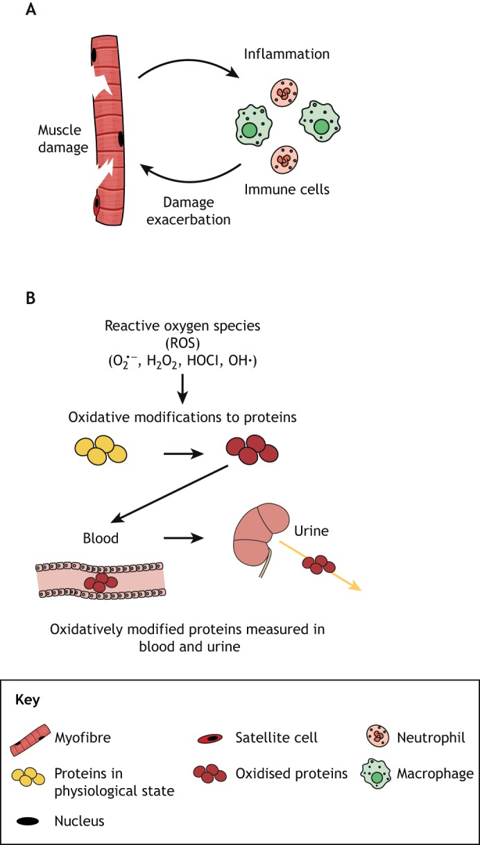
, dismutation of which leads to the formation of hydrogen peroxide (H2O2) that is either catalysed by MPO to form the highly cytotoxic oxidant hypochlorous acid (HOCl), or is further oxidised to generate hydroxyl radicals (OH•). These oxidants can potentially exacerbate necrosis of dystrophic myofibres by the reversible and irreversible damage modifications that affect the function of cellular proteins. These modified proteins can enter circulation and are often excreted, therefore the measurement of these modifications in plasma and urine can be used as biomarkers of inflammation and oxidative stress in the muscle.
Fig. 5.
Fig. 5.
Home blood and urine collection to measure biomarkers of dystropathology. Biomarkers that can be measured in a drop of blood collected from a finger prick onto a card for storage at room temperature can be readily collected in the home by patients or their family. Similarly, analytes of dystropathology in urine can potentially be measured using an absorbent strip that is dried and stored at room temperature. Home collection would augment clinical utility by facilitating tracking of biomarkers.

References

    1. Aartsma-Rus A. and Spitali P. (2015). Circulating biomarkers for Duchenne muscular dystrophy. J. Neuromuscul. Dis. 2, S49-S58. 10.3233/JND-150102 - DOI - PMC - PubMed
    1. Aartsma-Rus A., Ferlini A., McNally E. M., Spitali P., Sweeney H. L. and Workshop participants (2018). 226(th) ENMC International Workshop: Towards validated and qualified biomarkers for therapy development for Duchenne muscular dystrophy 20-22 January 2017, Heemskerk, The Netherlands. Neuromuscul. Disord. 28, 77-86. 10.1016/j.nmd.2017.10.002 - DOI - PMC - PubMed
    1. Allen D. G., Whitehead N. P. and Froehner S. C. (2016). Absence of Dystrophin disrupts skeletal muscle signaling: roles of Ca2+, reactive oxygen species, and nitric oxide in the development of muscular dystrophy. Physiol. Rev. 96, 253-305. 10.1152/physrev.00007.2015 - DOI - PMC - PubMed
    1. Amthor H., Egelhof T., McKinnell I., Ladd M. E., Janssen I., Weber J., Sinn H., Schrenk H.-H., Forsting M., Voit T. et al. (2004). Albumin targeting of damaged muscle fibres in the mdx mouse can be monitored by MRI. Neuromuscul. Disord. 14, 791-796. 10.1016/j.nmd.2004.08.004 - DOI - PubMed
    1. Armstrong A. E., Zerbes R., Fournier P. A. and Arthur P. G. (2011). A fluorescent dual labeling technique for the quantitative measurement of reduced and oxidized protein thiols in tissue samples. Free Radic. Biol. Med. 50, 510-517. 10.1016/j.freeradbiomed.2010.11.018 - DOI - PubMed

Publication types