Review
. 2020 Mar 26;9(4):280.
doi: 10.3390/antiox9040280.

The Role of Hydrogen Peroxide and Peroxiredoxins throughout the Cell Cycle

Affiliations
Review

The Role of Hydrogen Peroxide and Peroxiredoxins throughout the Cell Cycle

Sukyeong Heo et al. Antioxidants (Basel). .

Abstract

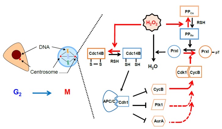
Hydrogen peroxide (H2O2) is an oxidizing agent that induces cellular damage at inappropriate concentrations and gives rise to an arrest during cell cycle progression, causing cell death. Recent evidence indicates that H2O2 also acts as a promoter for cell cycle progression by oxidizing specific thiol proteins. The intracellular concentration of H2O2 is regulated tightly, enabling its use as a cellular signaling molecule while minimizing its potential to cause cellular damage. Peroxiredoxins (Prxs) have peroxidase activity toward H2O2, organic hydroperoxides, and peroxynitrite for protecting cells from oxidative stress. They are suggested to work as signaling mediators, allowing the local accumulation of H2O2 by inactivating their peroxidase activity uniquely compared with other antioxidant proteins such as catalase and glutathione peroxidase. Given that Prxs are highly sensitive to oxidation by H2O2, they act as sensors and transducers of H2O2 signaling via transferring their oxidation state to effector proteins. The concentrations of intracellular H2O2 increase as the cell cycle progresses from G1 to mitosis. Here, we summarize the roles of Prxs with regard to the regulation of cell cycle-dependent kinase activity and anaphase-promoting complex/cyclosome in terms of changes in H2O2 levels. Protection of the cell from unwanted progression of the cell cycle is suggested to be a role of Prx. We discuss the possible roles of Prxs to control H2O2 levels.

Keywords: cell cycle; centrosome; cyclin-dependent kinase; hydrogen peroxide; peroxiredoxin.


Conflict of interest statement

The authors declare no conflict of interest.

Figures

Figure 1
Figure 1
The morphological change in subcellular compartments and the activities of cell cycle-dependent kinases (Cdks) and anaphase-promoting complex/cyclosome (APC/C) throughout the cell cycle in a mammalian cell: During the cell cycle, after DNA replication is finished at the S phase, a cell prepares to enter mitosis through the G2 phase. At the G2–M transition, the structure of subcellular organelles is changed dramatically as illustrated here. A nuclear envelop is broken and the Golgi (green), ER (yellow), and mitochondria (pink) are fragmented. After mitotic exit, all the organelles are reorganized and structured in filamentous states. Activity of APC/C is inversely propositioned to that of Cdks during cell cycle progression except at the G2 phase. APC/C activity is inhibited by early mitotic inhibitor1 at the G2 phase. In G0 (quiescent phase) cells, APC/C–Cdh1 remains active.
Figure 2
Figure 2
Localization of six peroxiredoxin isoforms (PrxI–PrxVI) inside and outside of the mammalian cell: In the cytosolic space, PrxI, II, IV, V, and VI are present. The nucleus has PrxI, PrxII, and PrxV, and mitochondria have PrxIII and PrxV. In the peroxisome, PrxV exists, and in the ER, PrxIV is present. The lysosome has PrxIV and PrxVI. PrxI is also associated with the centrosome. PrxIV and PrxVI are present outside of the cell.
Figure 3
Figure 3
Oscillation of H2O2 levels during the cell cycle (experimental data by the authors): (A,B) HeLa cells expressing cytosolic HyPer (cHyPer) were arrested at prometaphase by treatment with thymidine and nocodazole (T/N) and then released into G1 phase by shaking off into fresh medium and incubation for 3 h (T/N+R3). The fluorescence intensity of cHyPer was measured by flow cytometry, and the relative levels of H2O2 were estimated (A) for asynchronous, T/N, and T/N+R3 cells. Asynchronous HeLa cells that were not transfected with the cHyPer vector were analyzed similarly. Cell cycle status was also determined by flow cytometric analysis after staining with propidium iodide (B). Data in (A) are means ± SD from three independent experiments. (C) The relative fluorescence intensity of cHyPer–C199S was measured as in (A). Data are from a representative experiment. (D–F) HeLa cells expressing cHyPer were arrested at G1–S with a double-thymidine block and released into fresh medium. The relative levels of H2O2 were measured by flow cytometry at the indicated times after the release (D). Cell cycle status was also analyzed by flow cytometry after staining with propidium iodide (E), and the expression of cell cycle marker proteins was examined by immunoblot analysis (F). cHyPer is detected with antibodies to green fluorescent protein. Data are from a representative experiment. pHH3, phosphorylated histone H3. Rabbit polyclonal antibodies to Ser10-phosphorylated histone H3 (pHH3) (from Upstate Biotechnology) were used to detect mitotic cells. For analysis of cell cycle stage, cells (5 × 10⁵/mL) were washed twice with ice-cold phosphate-buffered saline, fixed overnight at 4 °C in 70% ethanol, and stained with 1 mL of a solution containing RNase (50 μg/mL) and propidium iodide (50 μg/mL) before flow cytometry with a FACSCalibur instrument (BD Biosciences, Franklin Lakes, USA). For measurement of the intracellular level of H2O2, cells expressing cHyPer or cHyPer-C199S were analyzed at an excitation wavelength of 488 nm.
Figure 4
Figure 4
Protective role of peroxiredoxin I (PrxI) in the cell under oxidative stress: Centrosome-associated PrxI proteins shield the organelles from a high tide of H2O2 molecules from various intracellular sources at G2 phase. Anaphase-promoting complex/cyclosome (APC/C)–Cdh1 activity is high at the centrosome, and therefore, its substrates such as cyclin B, polo-like kinase1 (Plk1), and Aurora A are degraded. During mitosis, PrxI is inactivated by cell cycle-dependent kinase 1 (Cdk1) through phosphorylation and the large numbers of H2O2 molecules inhibit APC/C-Cdh1, which results in increased cyclin B, Plk1, and Aurora A proteins. Centrosome-associated PrxI can protect cells from other oxidative stress, including ultraviolet (UV) or ionizing radiation (IR).
Figure 5
Figure 5
A model illustrating the role of pericentrosomal H2O2 for activation of cell cycle-dependent kinase 1 (Cdk1): Red arrows indicate the direction of the positive feedback reactions resulting from peroxiredoxin I (PrxI) phosphorylation by Cdk1 at the mitotic centrosomes. Black arrows indicate the direction of those resulting from PrxI dephosphorylation. Dashed red arrows indicate multiple positive feedback loops known to activate Cdk1 directly or indirectly. AurA, Aurora A; CycB, cyclin B; Plk1, polo-like kinase1; APC/C, anaphase-promoting complex/cyclosome; PP, protein phosphatase; Cdc14B, cell division cycle 14B. See Section 6 for details.

References

    1. Giorgio M., Trinei M., Migliaccio E., Pelicci P.G. Hydrogen peroxide: A metabolic by-product or a common mediator of ageing signals? Nat. Rev. Mol. Cell Biol. 2007;8:722–728. doi: 10.1038/nrm2240. - DOI - PubMed
    1. Rhee S.G. Cell signaling. H2O2, a necessary evil for cell signaling. Science. 2006;312:1882–1883. doi: 10.1126/science.1130481. - DOI - PubMed
    1. Suh Y.A., Arnold R.S., Lassegue B., Shi J., Xu X., Sorescu D., Chung A.B., Griendling K.K., Lambeth J.D. Cell transformation by the superoxide-generating oxidase Mox1. Nature. 1999;401:79–82. doi: 10.1038/43459. - DOI - PubMed
    1. van der Vliet A., Hristova M., Cross C.E., Eiserich J.P., Goldkorn T. Peroxynitrite induces covalent dimerization of epidermal growth factor receptors in A431 epidermoid carcinoma cells. J. Biol. Chem. 1998;273:31860–31866. doi: 10.1074/jbc.273.48.31860. - DOI - PubMed
    1. Clempus R.E., Griendling K.K. Reactive oxygen species signaling in vascular smooth muscle cells. Cardiovasc. Res. 2006;71:216–225. doi: 10.1016/j.cardiores.2006.02.033. - DOI - PMC - PubMed

LinkOut - more resources