. 2020 Mar 26;21(7):2292.
doi: 10.3390/ijms21072292.

The Expression of Selected Factors Related to T Lymphocyte Activity in Canine Mammary Tumors

Affiliations

The Expression of Selected Factors Related to T Lymphocyte Activity in Canine Mammary Tumors

Joanna K Bujak et al. Int J Mol Sci. .

Abstract

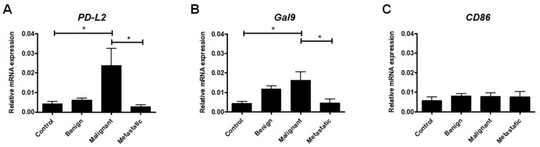
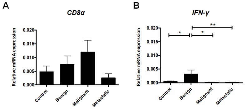
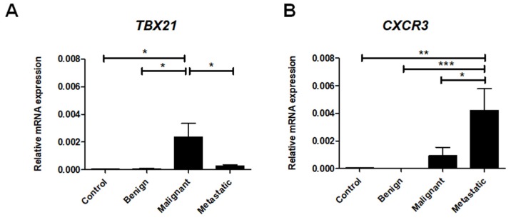
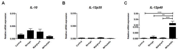
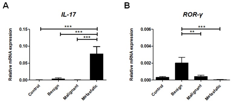
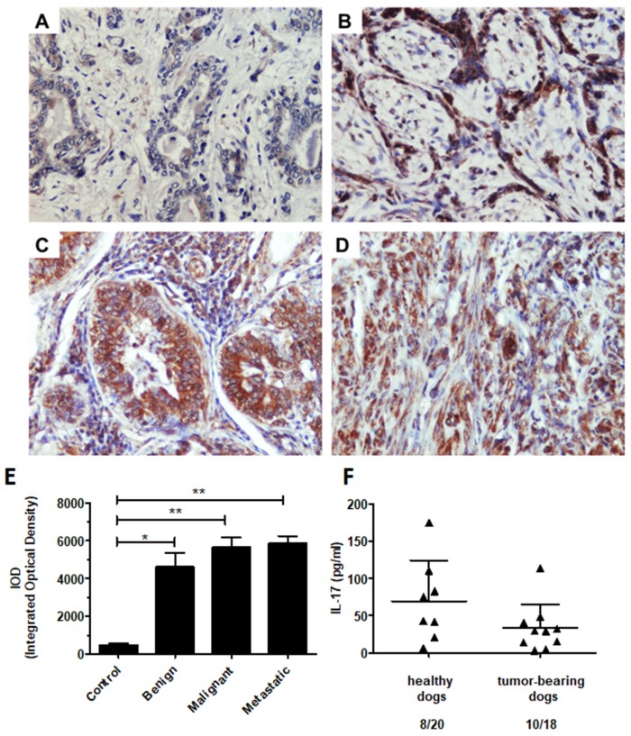
Crosstalk between neoplastic and immune cells in the tumor microenvironment (TME) influences the progression of disease in human and canine cancer patients. Given that canine mammary tumors are a useful model to study breast cancer biology, we aimed to evaluate the expression of genes associated with T lymphocyte activity in benign, malignant, and metastatic canine mammary tumors. Interestingly, metastatic tumors exhibit increased expression of CXCR3, CCR2, IL-4, IL-12p40, and IL-17. In particular, we focused on IL-17, a key interleukin associated with the Th17 lymphocyte phenotype. Th17 cells have been shown to play a contradictory role in tumor immunity. Although IL-17 showed a high expression in the metastatic tumors, the expression of RORγt, a crucial transcription factor for Th17 differentiation was barely detected. We further investigated IL-17 expression using immunohistochemistry, through which we confirmed the increased expression of this interleukin in malignant and metastatic mammary tumors. Finally, we compared the plasma levels of IL-17 in healthy and malignant mammary tumor-bearing dogs using ELISA but found no differences between the groups. Our data indicate that the IL-17 in metastatic tumors may be produced by other cell types, but not by Th17 lymphocytes. Overall, our results broaden the available knowledge on the interactions in canine mammary tumors and provide insight into the development of new therapeutic strategies, with potential benefits for human immune oncology.

Keywords: Th17 cells; breast cancer model; cancer immunotherapy; chemokine receptors; co-inhibitory ligands; immune checkpoints; inflammatory cytokines; interleukin-17; tumor-associated macrophages.


Conflict of interest statement

The authors declare no conflicts of interest. The funders had no role in the design of the study; in the collection, analyses, or interpretation of data; in the writing of the manuscript, or in the decision to publish results.

Figures

Figure 1
Figure 1
The expression of ligands regulating T cell activation at the mRNA level in canine mammary tumors. The relative PD-L2 (A), Gal9 (B), and CD86 (C) gene expression levels in healthy mammary gland tissue (Control) and in benign, malignant, and metastatic canine mammary tumors. The results are presented as the mean ± SEM. A one-way ANOVA and Tukey HSD post hoc test were applied. Significance levels are indicated as follows: * p < 0.05.
Figure 2
Figure 2
Relative gene expression of CD8α (A) and IFN-γ (B) in healthy mammary gland tissue (Control) and in benign, malignant, and metastatic canine mammary tumors. The results are presented as the mean ± SEM. A one-way ANOVA and Tukey HSD post hoc test were applied. Significance levels are indicated as follows: * p < 0.05; ** p < 0.01; *** p < 0.001.
Figure 3
Figure 3
Th1 cell-associated gene expression levels in canine mammary tumors. The relative gene expression of CXCR3 (A) and TBX21 (B) in healthy mammary gland tissue (Control) and in benign, malignant, and metastatic canine mammary tumors. The results are presented as the mean ± SEM. A one-way ANOVA and Tukey HSD post hoc test were applied. Significance levels are indicated as follows: * p < 0.05; ** p < 0.01; *** p < 0.001.
Figure 4
Figure 4
Th2 cell-associated gene expression levels in canine mammary tumors. The relative gene expression of CCR2 (A), GATA3 (B), and IL-4 (C) in healthy mammary gland tissue (Control) and in benign, malignant, and metastatic canine mammary tumors. The results are presented as the mean ± SEM. A one-way ANOVA and Tukey HSD post hoc test were applied. Significance levels are indicated as follows: * p < 0.05; ** p < 0.01; *** p < 0.001.
Figure 5
Figure 5
The expression of inflammatory cytokines at the mRNA level in canine mammary tumors. Relative gene expression of IL-10 (A), IL-12p35 (B), and IL-12p40 (C) in healthy mammary gland tissue (Control) and in benign, malignant, and metastatic canine mammary tumors. The results are presented as the mean ± SEM. A one-way ANOVA and Tukey HSD post hoc test were applied. Significance levels are indicated as follows: ** p < 0.01; *** p < 0.001.
Figure 6
Figure 6
Th17 cell-associated gene expression levels in canine mammary tumors. Relative gene expression of ROR-γt (A) and IL-17 (B) in healthy mammary gland tissue (Control) and in benign, malignant, and metastatic canine mammary tumors. The results are presented as the mean ± SEM. A one-way ANOVA and Tukey HSD post hoc test were applied. Significance levels are indicated as follows: * p < 0.05; ** p < 0.01; *** p < 0.001.
Figure 7
Figure 7
The immunohistochemical expression of IL-17 in canine mammary tumors and the plasma levels of IL-17 in healthy and mammary tumor-bearing dogs. Representative light micrographs of canine mammary gland tissue (A), benign (B), malignant (C), and metastatic tumors (D) obtained with an Olympus BX60 microscope (at 200× total magnification). The IL-17 antigen is represented by the brown-colored precipitate in the cell cytoplasm and extracellular matrix. (E) The graph showing the IOD (integrated optical density) of IL-17 expression. The results are presented as the mean ± SEM. A one-way ANOVA and Tukey HSD post hoc test were applied. Significance levels are indicated as follows: * p < 0.05; ** p < 0.01. (F) The concentration of IL-17A (pg/mL) in the plasma of the client-owned healthy (8 out of 21) and malignant canine mammary tumor-bearing dogs (10 out of 18). Student’s t- test was applied, and no statistical significance was observed (p > 0.05).

References

    1. Nunes F.C., Damasceno K.A., De Campos C.B., Bertagnolli A.C., Lavalle G.E., Cassali G.D. Mixed tumors of the canine mammary glands: Evaluation of prognostic factors, treatment, and overall survival. Vet. Anim. Sci. 2019;7:100039. doi: 10.1016/j.vas.2018.09.003. - DOI - PMC - PubMed
    1. Choi J.-W., Yoon H.-Y., Jeong S.-W. Clinical Outcomes of Surgically Managed Spontaneous Tumors in 114 Client-owned Dogs. Immune Netw. 2016;16:116–125. doi: 10.4110/in.2016.16.2.116. - DOI - PMC - PubMed
    1. Philibert J.C., Snyder P.W., Glickman N., Glickman L.T., Knapp D.W., Waters D.J. Influence of host factors on survival in dogs with malignant mammary gland tumors. J. Vet. Intern. Med. 2003;17:102–106. doi: 10.1111/j.1939-1676.2003.tb01330.x. - DOI - PubMed
    1. Abdelmegeed S.M., Mohammed S. Canine mammary tumors as a model for human disease. Oncol. Lett. 2018;15:8195–8205. doi: 10.3892/ol.2018.8411. - DOI - PMC - PubMed
    1. Park J.S., Withers S.S., Modiano J.F., Kent M.S., Chen M., Luna J.I., Culp W.T.N., Sparger E.E., Rebhun R.B., Monjazeb A.M., et al. Canine cancer immunotherapy studies: Linking mouse and human. J. Immunother. Cancer. 2016;4:97. doi: 10.1186/s40425-016-0200-7. - DOI - PMC - PubMed

MeSH terms

LinkOut - more resources