Review
. 2008 Nov;252(21):2239-2277.
doi: 10.1016/j.ccr.2008.01.016. Epub 2008 Jan 26.

Applications of heteronuclear NMR spectroscopy in biological and medicinal inorganic chemistry

Affiliations
Review

Applications of heteronuclear NMR spectroscopy in biological and medicinal inorganic chemistry

Luca Ronconi et al. Coord Chem Rev. 2008 Nov.

Abstract

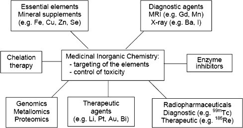
There is a wide range of potential applications of inorganic compounds, and metal coordination complexes in particular, in medicine but progress is hampered by a lack of methods to study their speciation. The biological activity of metal complexes is determined by the metal itself, its oxidation state, the types and number of coordinated ligands and their strength of binding, the geometry of the complex, redox potential and ligand exchange rates. For organic drugs a variety of readily observed spin I = 1/2 nuclei can be used (1H, 13C, 15N, 19F, 31P), but only a few metals fall into this category. Most are quadrupolar nuclei giving rise to broad lines with low detection sensitivity (for biological systems). However we show that, in some cases, heteronuclear NMR studies can provide new insights into the biological and medicinal chemistry of a range of elements and these data will stimulate further advances in this area.

Keywords: ADP, adenosine diphosphate; AES, atomic emission spectroscopy; AMP, adenosine monophosphate; ATP, adenosine triphosphate; BNCT, boron neutron capture therapy; BPG, 2,3-bisphosphoglycerate; BSA, bovine serum albumin; BSH, sodium borocaptate; Bioinorganic chemistry; Biological systems; DNA, deoxyribonucleic acid; EDTA-N4, ethylenediaminetetraacetamide; EFG, electric field gradient; GMP, guanosine monophosphate; HMQC, heteronuclear multiple quantum correlation; Heteronuclear NMR spectroscopy; Im, imidazole; In, indazole; MQF, multiple quantum filtered; MRI, magnetic resonance imaging; Medicinal inorganic chemistry; Metallopharmaceuticals; NOE, nuclear Overhauser effect; PET, positron emission tomography; Quadrupolar nuclei; RBC, red blood cell; RNA, ribonucleic acid; SDS, sodium dodecyl sulfate; rRNA, ribosomal ribonucleic acid; tRNA, transfer ribonucleic acid.


Figures

Fig. 1
Fig. 1
Some of the areas of medicinal inorganic chemistry (adapted from Ref. [160]).
Fig. 2
Fig. 2
7Li NMR spectrum of a blood sample treated with Li2CO3 recorded in 30 min using a home-built solenoid coil at 7 T field strength. The chemical shift of lithium accumulated in RBCs has been set to 0 ppm. The RBC and plasma 7Li resonances are separated by 13 ppm by the addition of 20 mg of [Tm(DOTP)]5− as shift reagent (adapted from Ref. [29]).
Fig. 3
Fig. 3
(a) Variable-temperature 23Na NMR spectra of 0.80 M Na2(5′-GMP) obtained on a Bruker Avance-500 spectrometer (B0 = 11.75 T) operating at 132.26 MHz for 23Na. (b) 23Na NMR spectra of d(TG4T) obtained on a Bruker Avance-600 spectrometer (B0 = 14.1 T). An inversion-recovery sequence was used to partially suppress the large signal from the free Na+ ions. A recovery delay of 13 ms (close to the null point) was used. Typically, 250k scans were accumulated with a recycle time of 50 ms. The d(TG4T) DNA sample was prepared at 8 mM strand concentration in 10 mM sodium phosphate buffer (pH 7.1) and 100 mM NaCl. All 23Na chemical shifts are referenced to Na+(aq) at 0.0 ppm (adapted from Ref. [36]).
Fig. 4
Fig. 4
Variable-temperature 39K NMR spectra of 0.53 M Na2(5′-GMP) in presence of 0.10 M K+ obtained on a Bruker Avance-500 spectrometer (B0 = 11.75 T) operating at 23.33 MHz for 39K nucleus. Typically, 500k scans were accumulated with a recycle time of 10 ms. All 39K chemical shifts are referenced to K+(aq) at 0.0 ppm (adapted from Ref. [36]).
Fig. 5
Fig. 5
39K NMR spectrum of human RBCs resuspended in the NMR buffer at 37 °C. Ki+ and Ko+ represent the intra- and extracellular 39K peaks, respectively. Hematocrit was 61.2%. One division in the spectrum is equal to 50 ppm (adapted from Ref. [42]).
Fig. 6
Fig. 6
87Rb NMR spectra (referenced to internal Rb+(aq) at 0.0 ppm) of perfused rat heart during Rb+ load and washout. The hearts were perfused with phosphate-free Krebs–Henseleit buffer. The perfusate was equilibrated with 95% O2/5% CO2, with the pH maintained at 7.4. The buffer used for Rb+ loading contained 0.94 mM Rb+ and 3.76 mM K+ (adapted from Ref. [45]).
Fig. 7
Fig. 7
133Cs NMR spectra of human erythrocytes suspended in a buffer containing 140 mM NaCl and 10 mM CsCl. The origin of the chemical shift scale is arbitrary (adapted from Ref. [49]).
Fig. 8
Fig. 8
43Ca NMR spectra of the titration of 0.33 mM equine apolysozyme with 43Ca2+ at pH 6.0. 200k scans were collected in each experiment: (a) 0.23 equiv. of 43Ca2+; (b) 0.46 equiv. of 43Ca2+; (c) 0.69 equiv. of 43Ca2+; (d) 0.92 equiv. of 43Ca2+; (e) 1.15 equiv. of 43Ca2+ (adapted from Ref. [68]).
Fig. 9
Fig. 9
Schematic illustration of the BSH anion. There is a net negative charge of 2 in the boron-hydride cage. δ(11B) of free BSH are referenced to saturated H3BO3 at 0.0 ppm (adapted from Ref. [73]).
Fig. 10
Fig. 10
The 11B (a) single quantum and (b) MQF (τ = 1.4 ms) NMR spectra of 100 mM borate solution with 4 mM ferricytochrome-c at 5 °C, 11.4 T, and pH 9.7 (adapted from Ref. [75]).
Fig. 11
Fig. 11
Backbone structure of human serum transferrin (coordinates supplied by Zuccola [245]); only the C-lobe site is occupied by iron in this structure.
Fig. 12
Fig. 12
27Al NMR spectra (referenced to external 1.0 M [Al(NO3)3] in D2O at 0.0 ppm) of ovotransferrin (oTf, 1.13 mM, pH 7.5), serotransferrin (sTr, 1.09 mM, pH 7.3), and lactoferrin (lTf, 0.73 mM, pH 7.5) in the presence of 20 mM Na213CO3 and 2.0 equiv. of Al3+ (75% H2O/25% D2O, 150 mM KCl, 25 °C) at four magnetic fields: 7.0 T (νo = 78.2 MHz), 9.4 T (νo = 104.3 MHz), 11.7 T (νo = 130.3 MHz), and 14.1 T (νo = 156.4 MHz) (adapted from Ref. [82]).
Fig. 13
Fig. 13
27Al NMR spectra (referenced to external 1.0 M [Al(NO3)3] in D2O at 0.0 ppm) of (a) 1.20 mM ovotransferrin in the presence of 5 mM Na213C2O4 and 2.0 equiv. of Al3+ (75% H2O/25% D2O, 150 mM KCl, pH 7.4, 25 °C), and (b) 1.13 mM ovotransferrin in the presence of 20 mM Na213CO3 and 2.0 equiv. of Al3+ (75% H2O/25% D2O, 150 mM KCI, pH 7.6, 25 °C) at a magnetic field strength of 11.7 T (130.3 MHz) (adapted from Ref. [81a]).
Fig. 14
Fig. 14
207Pb (104.435 MHz) NMR spectra of the 207Pb2+ forms of bacterially expressed mammalian calmodulin (1.47 mM, 4.0 equiv. 207Pb2+, 100 mM KCl, pH 7.1, 85k scans) and its C-terminal domain fragment TR2C (1.11 mM, 2.0 equiv. 207Pb2+, 100 mM KCl, pH 7.0, 80k scans) (adapted from Ref. [114]).
Fig. 15
Fig. 15
2D [1H,207Pb] HMQC NMR spectrum of [207Pb(EDTA-N4)]2+ (Pb-ethylenediaminetetraacetamide), 99.9% D2O, pH 6.1, 25 °C). Coupling is observed from 207Pb to 1H through three bonds. The lead chemical shift was referenced to external 1 M [Pb(NO3)2] in 99.9% D2O at pH 3.3 (adapted from Ref. [112]).
Fig. 16
Fig. 16
13C and 45Sc NMR spectra (referenced to external neat TMS and 1.0 M ScCl3 in D2O, respectively, at 0.0 ppm) of 1.05 mM ovotransferrin in the presence of 10 mM Na213CO3 and 1.9 equiv. of Sc3+ (75% H2O/25% D2O, 150 mM KCI, pH 7.6, 25 °C) (adapted from Ref. [82c]).
Fig. 17
Fig. 17
Variable pH 51V NMR spectra (referenced to external neat [VOCl3] at 0.0 ppm) of a 0.15 M NaCl aqueous solution of (a) [VIVO(maltolato)2] (10 mM) and (b) NH4[VVO2(maltolato)2] (10 mM) at 25 °C (adapted from Ref. [136]).
Fig. 18
Fig. 18
57Fe NMR spectra (referenced to external [Fe(CO)5] at 0.0 ppm) of 57Fe-cytochrome-c (57Fe-Cyt-c, 3 mM in 50 mM phosphate buffer, pH 7, 298 K), 57Fe-carbonmonoxymyoglobin (57Fe-MbCO, 15 mM in 50 mM phosphate buffer, pH 7.1, 296 K), and ferrocene (Cp2Fe, 0.8 M in C6H6) (adapted from Refs. [158c,158d]).
Fig. 19
Fig. 19
Structure of vitamin B12.
Fig. 20
Fig. 20
59Co NMR spectra of natural B12 derivatives in solution (externally referenced to 1 M aqueous [Co(NH3)6]Cl3, and subsequently converted to ppm downfield from 1 M aqueous K3[Co(CN)6] for the sake of literature consistency). Superimposed on the experimental traces are best Lorentzian fits characterized by the isotropic shift δ and full-width-at-half-height (FWHH) values indicated on the right (adapted from Ref. [174]).
Fig. 21
Fig. 21
2D [1H,109Ag] HMQC spectrum (referenced to external 1 M AgNO3 at 0.0 ppm) of yeast 109Ag-metallothionein (6 mM in 18 mM phosphate buffer in D2O, pD 6.5, 298 K). The spectrum shows the connectivities of all seven silver atoms with the cysteine Hβ protons of the yeast Ag-metallothionein (adapted from Ref. [190]).
Fig. 22
Fig. 22
Observed 113Cd chemical shifts for structurally characterized 113Cd-substituted metalloproteins: mononuclear (top) and polynuclear (bottom). All shifts are reported relative to external 0.1 M [Cd(ClO4)2]. The chemical shift position is correlated to ligand composition, where S represents sulfur from cysteine, S* represents sulfur from methionine, O represents oxygen from carboxylate or water, and N represents nitrogen from histidine (adapted from Ref. [201a]).
Fig. 23
Fig. 23
113Cd NMR spectra of mammalian MTs (mouse MT-1, human MT-3, rabbit MT-2) acquired at the indicated frequencies. Schematic structures of the two metal clusters are reported (adapted from Ref. [201a]).
Fig. 24
Fig. 24
Three-dimensional coronal 17O MRI (Fourier series window) images of natural abundance H217O in the rat brain acquired at 9.4 T. Three adjacent coronal 17O images (0.1 ml voxel size; total acquisition time of 11 s) and the corresponding proton anatomical images are presented herein (adapted from Ref. [220a]).
Fig. 25
Fig. 25
In vivo33S NMR spectrum (referred to the 33S signal of 1 M Na2SO4 in H2O in a coaxial cell) of an L. lithophaga homogenate. The peak at −6.8 ppm is assigned to the amino acid taurine (adapted from Ref. [223]).
Fig. 26
Fig. 26
19F NMR spectrum at 282 MHz with proton decoupling of a urine sample from a patient treated orally with capecitabine at a dose of 2900 mg/day administered twice daily at 12 h interval. Urine fraction 0–12 h collected after the first dose of 1450 mg and 14-fold concentrated, pH of the sample: 5.5. The chemical shifts are expressed relative to the resonance peak of trifluoroacetic acid (5% (w/v) aqueous solution) used as external reference (REF). Assignments: F, fluoride ion; CAP, capecitabine; 5′dFCR, 5′-deoxy-5-fluorocytidine; 5′dFUR, 5′-deoxy-5-fluorouridine; FC, 5-fluorocytosine; FU, 5-fluorouracil; ?, unknown; FUPA, α-fluoro-β-ureidopropionic acid; FBAL, α-fluoro-β-alanine; FHPA, 2-fluoro-3-hydroxypropanoic acid; OHFC, 6-hydroxy-5-fluorocytosine; FUH2, 5,6-dihydro-5-fluorouracil; FAC, fluoroacetic acid. J, 1J(13C–19F) coupling constant (adapted from Ref. [16h]).
Fig. 27
Fig. 27
Catabolic pathway of capecitabine. All the compounds (except CFBAL) are represented in neutral form. CAP, capecitabine; 5′dFCR, 5′-deoxy-5-fluorocytidine; 5′dFUR, 5′-deoxy-5-fluorouridine; FU, 5-fluorouracil; FUH2, 5,6-dihydro-5-fluorouracil; FUPA, α-fluoro-β-ureidopropionic acid; FBAL, α-fluoro-β-alanine; CFBAL, N-carboxy-α-fluoro-β-alanine; F, fluoride ion; FHPA, 2-fluoro-3-hydroxypropanoic acid; FAC, fluoroacetic acid; FC, 5-fluorocytosine; OHFC, 6-hydroxy-5-fluorocytosine.

References

    1. McMaster J. Annu. Rep. Prog. Chem. Sect. A: Inorg. Chem. 2002;98:593.
    1. Sadler P.J., Muncie C., Shipman M.A. In: Biological Inorganic Chemistry: Structure & Reactivity. Bertini I., Gray H.B., Stiefel E.I., Valentine J.S., editors. University Science Books; Mill Valley, CA: 2007. p. 95.
    1. Park M., Li Q., Shcheynikov N., Muallem S., Zeng W. Cell Cycle. 2005;4:24. - PubMed
    1. Camilleri C., Markich S.J., Noller B.N., Turley C.J., Parker G., van Dam R.A. Chemosphere. 2002;50:355. - PubMed
    1. Levina A., Lay P.A. Coord. Chem. Rev. 2005;249:281.

LinkOut - more resources