. 2020 Mar 12:11:296.
doi: 10.3389/fphar.2020.00296. eCollection 2020.

Novel Anti-Interleukin-1β Therapy Preserves Retinal Integrity: A Longitudinal Investigation Using OCT Imaging and Automated Retinal Segmentation in Small Rodents

Affiliations

Novel Anti-Interleukin-1β Therapy Preserves Retinal Integrity: A Longitudinal Investigation Using OCT Imaging and Automated Retinal Segmentation in Small Rodents

Diane N Sayah et al. Front Pharmacol. .

Abstract

Retinopathy of prematurity (ROP) is the leading cause of blindness in neonates. Inflammation, in particular interleukin-1β (IL-1β), is increased in early stages of the disorder, and contributes to inner and outer retinal vasoobliteration in the oxygen-induced retinopathy (OIR) model of ROP. A small peptide antagonist of IL-1 receptor, composed of the amino acid sequence, rytvela, has been shown to exert beneficial anti-inflammatory effects without compromising immunovigilance-related NF-κB in reproductive tissues. We conducted a longitudinal study to determine the efficacy of "rytvela" in preserving the integrity of the retina in OIR model, using optical coherence tomography (OCT) which provides high-resolution cross-sectional imaging of ocular structures in vivo. Sprague-Dawley rats subjected to OIR and treated or not with "rytvela" were compared to IL-1 receptor antagonist (Kineret). OCT imaging and custom automated segmentation algorithm used to measure retinal thickness (RT) were obtained at P14 and P30; gold-standard immunohistochemistry (IHC) was used to confirm retinal anatomical changes. OCT revealed significant retinal thinning in untreated animals by P30, confirmed by IHC; these changes were coherently associated with increased apoptosis. Both rytvela and Kineret subsided apoptosis and preserved RT. As anticipated, Kineret diminished both SAPK/JNK and NF-κB axes, whereas rytvela selectively abated the former which resulted in preserved monocyte phagocytic function. Altogether, OCT imaging with automated segmentation is a reliable non-invasive approach to study longitudinally retinal pathology in small animal models of retinopathy.

Keywords: anti-interleukin-1β; automated segmentation; kineret; optical coherence tomography; oxygen-induced retinopathy; retina; rytvela; therapy.


Figures

FIGURE 1
FIGURE 1
Description of automated segmentation algorithm. Left: Identification of the sclera, the outermost retinal layer (ORL), and the innermost retinal layer (IRL) in a section of a typical rodent eye B-scan. Center: Intensity profile along the center A-scan of the image at the left, averaged with a Gaussian filter (σ = 20 μm). The absolute maximum signals the location of the sclera. Right: First derivative (slope) of the intensity profile along the center A-scan of the image at the left. The first valley above the sclera indicates the ORL, and the highest peak more than 120 μm above it indicates the IRL. The slope is computed after a Gaussian smoothing operation with σ = 2 μm.
FIGURE 2
FIGURE 2
Early anti-IL-1β therapy preserves retinal integrity in OIR subjects. (A) Original B-scans overlaid with the innermost layer of the retina (purple), the inner choroid limit (green), and the choroid-sclera interface (yellow) are shown for each sub-group at P30. (B) There is no difference in retinal thickness on P14. (C) However, OIR retinae become statistically thinner by P30; retinal thickness is preserved by anti-IL-1β. N = 4 animals on P14; N = 3-4 on P30. Values are mean ± SEM. One-way ANOVA; p < 0.05, ∗∗p < 0.01, ∗∗∗p < 0.001, ****p < 0.0001 vs corresponding values as indicated.
FIGURE 3
FIGURE 3
Early anti-IL-1 β therapy reduces the number of apoptotic cells and preserves retinal vessel density at the superficial capillary plexus of OIR subjects. (A–D) TUNEL staining show 2–4 positive cells per 2500 mm2 in P14 control (NOR) animals (with or without anti-IL-1β), compared to ∼10 cells in OIR rats of the same age. (E,F) Both IL-1β antagonists effectively diminish TUNEL positive cells in OIR subjects. Statistical analyses are shown in G. (H) P14 control animals display normal vessel density (green) in the retina. (I) P14 OIR rats show reduced vessel density at the superficial plexus. (J–L) Rytvela and Kineret preserved retinal vessels at the superficial capillary plexus of OIR rats. N = 3–4 animals per group. Values are mean ± SEM. One-way ANOVA; p < 0.05, ∗∗p < 0.01, ∗∗∗p < 0.001, ****p < 0.0001 vs corresponding values as indicated.
FIGURE 4
FIGURE 4
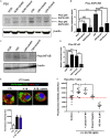
Different inhibition profiles between rytvela and Kineret in retinal tissue at P30 in OIR subjects vs. controls (NOR) and the effect of rytvela on macrophage phagocytosis. (A,B) Both rytvela and Kineret abolish SAPK/JNK phosphorylation in OIR subjects. (C,D) However, Kineret inhibits NF-κB, whereas rytvela preserves NF-κB pathway. N = 3–5 animals. Values are mean ± SEM; p < 0.05, ∗∗p < 0.01, ∗∗∗p < 0.001, ****p < 0.0001 vs corresponding values as indicated. (E) Confocal imaging showing Fluorescein-labeled BioParticles (green) phagocytosed by J774 mononuclear cells counterstained with Rhodamine phalloidin (red) and DAPI (blue). Histogram below immunofluorescent images refer to quantitative analysis using fluorescence intensity plate reading, showing that rytvela does not inhibit IL-1β-induced phagocytosis. (F) Quantitative analysis of Raw 264.7 cell phagocytosis activated by IL-1β, showing preservation by rytvela (1 μM) but not by Kineret (1 mg/ml) or the selective NF-kB inhibitor SC-514 (2 μM). N = 4–11/group. One-way ANOVA; p < 0.05, ∗∗p < 0.01, ∗∗∗p < 0.001, ****p < 0.0001 compared to control.

References

    1. Beaton L., Mazzaferri J., Lalonde F., Hidalgo-Aguirre M., Descovich D., Lesk M. R., et al. (2015). Non-invasive measurement of choroidal volume change and ocular rigidity through automated segmentation of high-speed OCT imaging. Biomed. Opt. Exp. 6 1694–1706. 10.1364/BOE.6.001694 - DOI - PMC - PubMed
    1. Beaudry-Richard A., Nadeau-Vallee M., Prairie E., Maurice N., Heckel E., Nezhady M., et al. (2018). Antenatal IL-1-dependent inflammation persists postnatally and causes retinal and sub-retinal vasculopathy in progeny. Sci. Rep. 8:11875. 10.1038/s41598-018-30087-30084 - DOI - PMC - PubMed
    1. Becker S., Wang H., Stoddard G. J., Hartnett M. E. (2017). Effect of subretinal injection on retinal structure and function in a rat oxygen-induced retinopathy model. Mol. Vis. 23 832–843. - PMC - PubMed
    1. Corson T. W., Samuels B. C., Wenzel A. A., Geary A. J., Riley A. A., McCarthy B. P., et al. (2014). Multimodality imaging methods for assessing retinoblastoma orthotopic xenograft growth and development. PLoS One 9:e99036. 10.1371/journal.pone.0099036 - DOI - PMC - PubMed
    1. Dorfman A., Dembinska O., Chemtob S., Lachapelle P. (2008). Early manifestations of postnatal hyperoxia on the retinal structure and function of the neonatal rat. Invest. Ophthalmol. Vis. Sci. 49 458–466. 10.1167/iovs.07-0916 - DOI - PubMed