Review
. 2019;7(9):25.
doi: 10.1007/s40134-019-0338-4. Epub 2019 Jul 11.

Imaging of Emerging Infectious Diseases

Affiliations
Review

Imaging of Emerging Infectious Diseases

Meghan Jardon et al. Curr Radiol Rep. 2019.

Abstract

Purpose of review: Emerging infectious diseases have seen a record increase in prevalence, and understanding their management is critical in an increasingly global community. In this paper, we review current literature detailing the role of radiology in the diagnosis and treatment of the Ebola (EVD), Zika (ZVD), Chikungunya (CHIKF), H1N1, Middle East Respiratory (MERS), and Severe Acute Respiratory Syndrome (SARS) viruses.

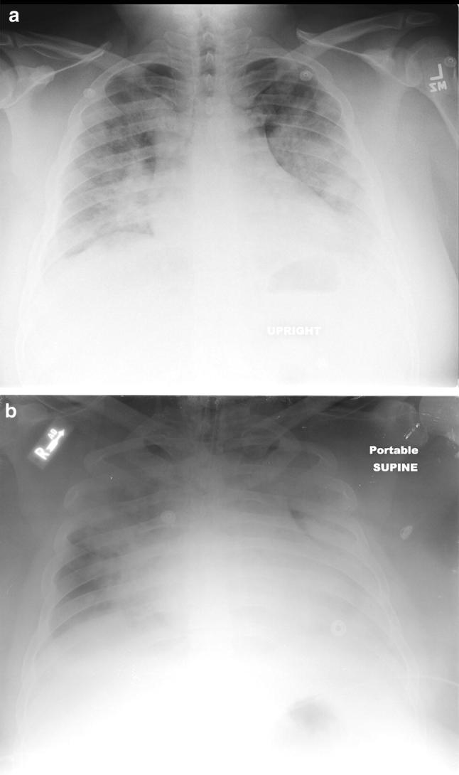
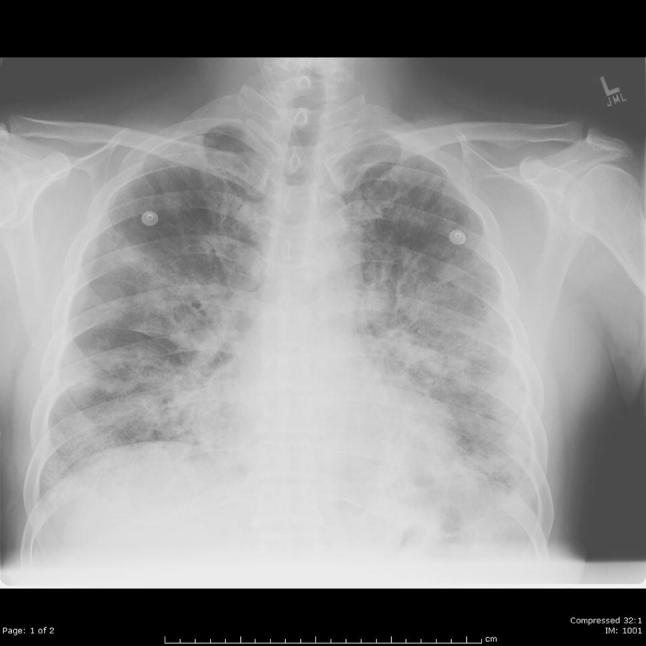
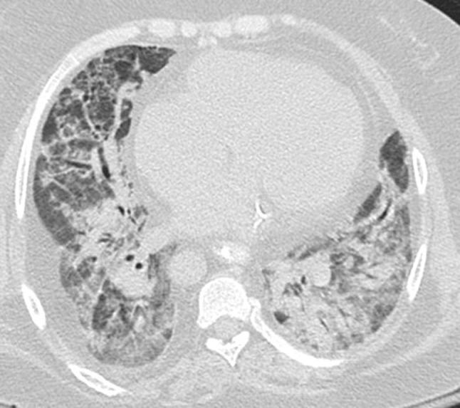
Recent findings: Complex protocols are required to safely use portable imaging in EVD to prevent nosocomial spread of disease. In ZVD, antenatal ultrasound can detect fetal abnormalities early, allowing implementation of care and support to affected families. Imaging is useful in assessing the extent of involvement of chronic CHIKF and monitoring treatment effect. Chest radiography and CT play a more direct role in the diagnosis and monitoring of the viral infections with primarily respiratory manifestations (H1N1, MERS, and SARS).

Summary: Radiology plays a variable role in emerging infectious diseases, requiring an understanding of disease transmission and safe imaging practices, as well as imaging features that affect clinical management.

Keywords: Chikungunya; Ebola; Emerging disease; H1N1; MERS; SARS; Zika.


Conflict of interest statement

Conflict of interestAll the authors declared that they have no potential conflicts of interest.

Figures

Fig. 1
Fig. 1
Infant with confirmed congenital Zika virus infection, with diffuse cortical atrophy and hypoplasia of the corpus callosum, cerebellum, and brainstem. a Coronal fluid-attenuated T2, b sagittal fluid-attenuated T2, c axial fat-saturated T1 and d sagittal T2 weighted MRI demonstrate these findings Images courtesy of Drs. Nielsen and Adachi, UCLA Department of Pediatrics. Originally published in “Zika Virus Infection in Pregnant Women in Rio de Janeiro – Preliminary Report” [21], reprinted with permission from NEJM
Fig. 2
Fig. 2
Maculopapular rash of Chikungunya involving the right foot Image courtesy of Clarissa Canella, MD, Universidade Federal Fluminense, Rio De Janeiro, Brazil
Fig. 3
Fig. 3
A 22-year-old female with Chikungunya fever who presented with 6 weeks of polyarthritis and low-grade fever. a Longitudinal ultrasound of the posterior tibial tendon (green arrow) demonstrates fluid and synovial proliferation (yellow arrow), indicating tenosynovitis associated with late stage Chikungunya fever. b Longitudinal ultrasound of the 3rd metatarsophalangeal joint demonstrates similar synovial proliferation (yellow arrow). c Power Doppler images show increased flow, indicating synovitis of the 4th metatarsophalangeal joint Images courtesy of Clarissa Canella, MD, Universidade Federal Fluminense, Rio De Janeiro, Brazil (Color figure online)
Fig. 4
Fig. 4
Patient who presented for evaluation 4 months after onset of polyarthritis. a Axial T2 weighted MRI of the left wrist demonstrates synovitis of the radiocarpal and metacarpophalangeal joints, as well as tenosynovitis of multiple extensor and flexor tendons (blue stars). b Axial T2 weighted MRI of the left ankle in a patient with Chikungunya infection, 4 months after symptom onset, demonstrates tenosynovitis involving the posterior tibialis and flexor digitorum longus tendons (green arrows) Images courtesy of Clarissa Canella, MD, Universidade Federal Fluminense, Rio De Janeiro, Brazil
Fig. 5
Fig. 5
A 34-year-old male with H1N1 influenza with rapid progression of disease. a Chest radiograph on the day of admission shows bilateral, symmetric ground glass and consolidation with (b) rapid progression of bilateral pulmonary opacities on hospital day 3. The patient ultimately required intubation and ICU admission
Fig. 6
Fig. 6
A 45-year-old male with H1N1 influenza who required intubation. Focal consolidation in the right lower lobe on chest radiograph is consistent with superimposed bacterial pneumonia
Fig. 7
Fig. 7
A 52-year-old male with H1N1 influenza with bilateral ground glass and consolidation and reticular opacities on chest radiograph
Fig. 8
Fig. 8
A 42-year-old female who initially presented with fever and shortness of breath. a Axial and b coronal CT of the chest demonstrate diffuse peribronchovascular consolidation on CT, subsequently confirmed as H1N1 viral pneumonia
Fig. 9
Fig. 9
A 30-year-old female with end-stage renal disease status post renal transplant who presented with H1N1 pneumonia that rapidly progressed to hypoxic respiratory failure and ARDS
Fig. 10
Fig. 10
A 40-year-old female with multiple medical comorbidities who initially presented with fever and severe cough, subsequently confirmed as MERS. Chest radiograph on admission to the ICU showed extensive air space consolidation in the right mid and the lower lung zones with ground glass opacities in the left mid lung zone. This patient expired 5 days after admission Image courtesy of Dr. K.M. Das, MD, FSCCT, Department of Radiology, College of Medicine and Health Science, UAE University, United Arab Emirates

References

    1. World Health Organization. The World Health Report 2007: a safer future: global public health security in the 21st century, 2007, ISBN 978 92 4 156344 4. https://www.who.int/whr/2007/en/. Accessed 1 Feb 2019.
    1. The National Institute for Occupational Safety and Health. Emerging Infectious Diseases, 2018, Centers for Disease Control, https://www.cdc.gov/niosh/topics/emerginfectdiseases/default.html. Accessed 1 Feb 2019.
    1. Holmes EC, Dudas G, Rambaut A, Andersen KG. The evolution of Ebola virus: insights from the 2013-2016 epidemic. Nature. 2016;538(7624):193–200. doi: 10.1038/nature19790. - DOI - PMC - PubMed
    1. World Health Organization. Ebola Virus Disease, 2018. https://www.who.int/news-room/fact-sheets/detail/ebola-virus-disease. Accessed 1 Feb 2019.
    1. Centers for Disease Control. Ebola in Democratic Republic of the Congo, 2018. http://wwwnc.cdc.gov/trave/notices/alert/ebola-democratic-republic-of-th.... Accessed 1 Feb 2019.

LinkOut - more resources