

Primary Syphilis Presenting As a Chronic Lip Ulcer
- PMID: 32226687
- PMCID: PMC7096073
- DOI: 10.7759/cureus.7086
Primary Syphilis Presenting As a Chronic Lip Ulcer
Abstract
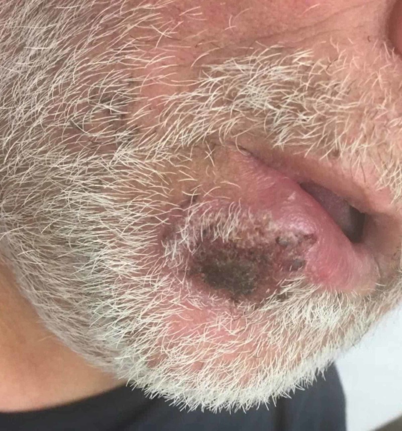
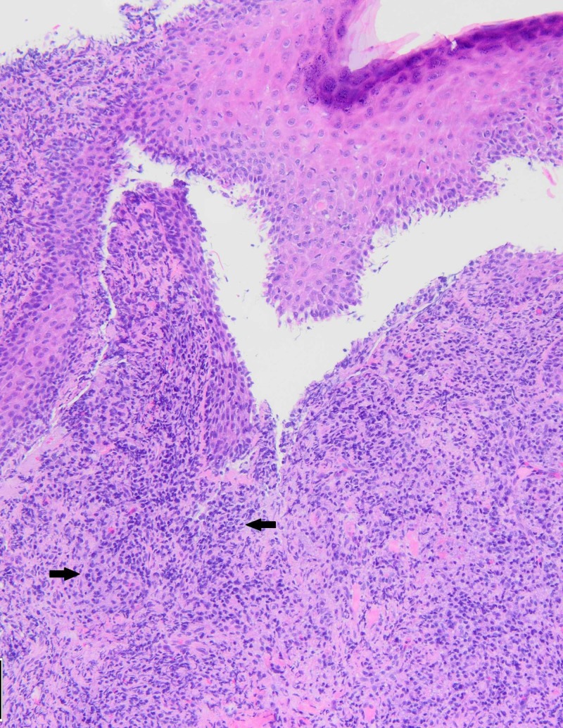
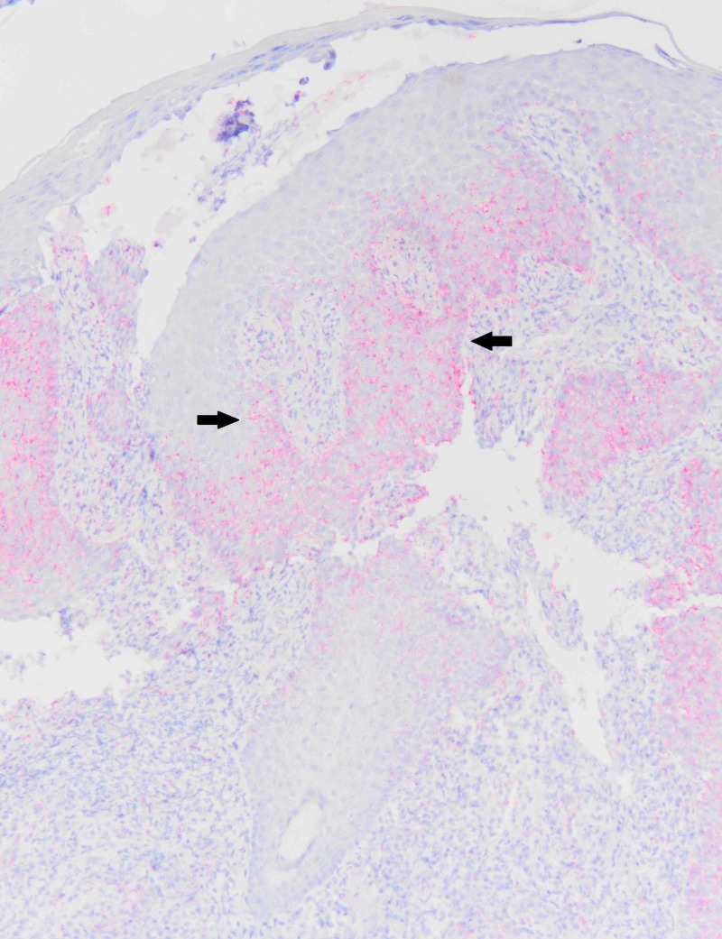
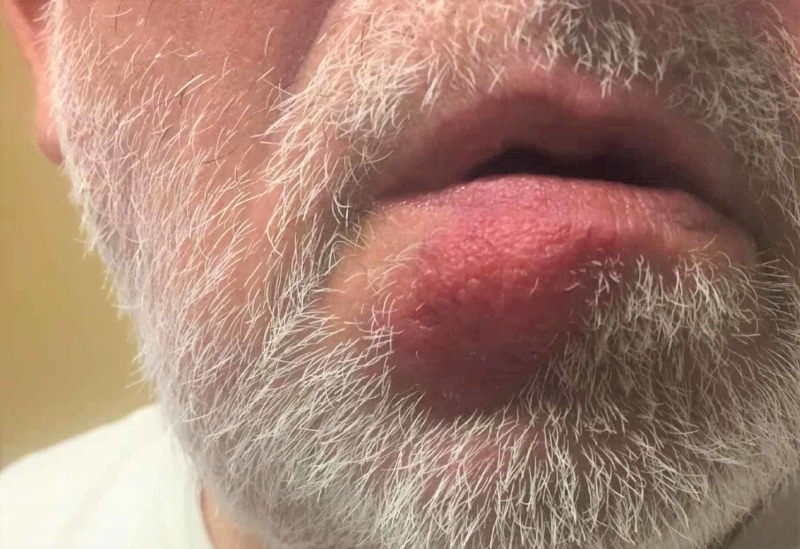
Syphilis is usually a sexually transmitted infection caused by the spirochete Treponema pallidum. Primary syphilis classically presents as a painless, ulcerated lesion on the genitals. However, the primary lesion is not restricted to this site and appears wherever the spirochete enters through the skin. The symptomatology and appearance of the primary lesion can also vary. We present a case of a 59-year-old man with a primary syphilitic chancre of the lower lip. The patient was referred to the dermatology clinic by their primary care provider after the ulceration failed to heal with antibiotic therapy. A biopsy of the lesion was taken at this time; the diagnosis of syphilis was then made by histologic examination and immunohistochemical staining. Subsequent serologic tests were also positive. Upon prompting, the patient did report a history of sexually transmitted disease but not of syphilis specifically. The patient was treated with penicillin, and there was clinical improvement of the lesion at the follow-up visit.
Keywords: biopsy; chancre; chronic ulcer; cutaneous; histology; oral syphilis; primary syphilis; skin; syphilis; treponema pallidum.
Copyright © 2020, Porterfield et al.
Conflict of interest statement
The authors have declared that no competing interests exist.
Figures
Similar articles
-
[Syphilitic chancre in the mouth: an unusual location. Case report].Rev Med Inst Mex Seguro Soc. 2022 Oct 25;60(6):703-707. Rev Med Inst Mex Seguro Soc. 2022. PMID: 36283073 Free PMC article. Spanish.
-
Sonographic appearance of syphilitic induration mimicking squamous cell carcinoma in the lower lip: a case report.J Med Case Rep. 2020 Nov 4;14(1):211. doi: 10.1186/s13256-020-02547-x. J Med Case Rep. 2020. PMID: 33143735 Free PMC article.
-
Multiple primary syphilis on the lip, nipple-areola and penis: An immunohistochemical examination of Treponema pallidum localization using an anti-T. pallidum antibody.J Dermatol. 2015 May;42(5):515-7. doi: 10.1111/1346-8138.12818. Epub 2015 Feb 24. J Dermatol. 2015. PMID: 25708895
-
Syphilitic Chancre of the Lips Transmitted by Kissing: A Case Report and Review of the Literature.Medicine (Baltimore). 2016 Apr;95(14):e3303. doi: 10.1097/MD.0000000000003303. Medicine (Baltimore). 2016. PMID: 27057901 Free PMC article. Review.
-
Primary syphilitic proctitis : case report and literature review.Acta Gastroenterol Belg. 2018 Jul-Sep;81(3):430-432. Acta Gastroenterol Belg. 2018. PMID: 30350534 Review.
References
-
- Clinical manifestations of primary syphilis. Bjekic M, Markovic M, Sipetic S. Braz J Infect Dis. 2012;16:387–389. - PubMed
-
- Neville BW, Damm DD, Allen CM, Chi A. Oral and Maxillofacial Pathology. Vol. 5. St. Louis: Elsevier; 2016. History of syphilis; pp. 164–190.
-
- Serologic response to treatment of infectious syphilis. Romanowski B, Sutherland R, Fick GH, Mooney D, Love EJ. Ann Intern Med. 1991;114:1005–1009. - PubMed
-
- Centers for Disease Control and Prevention. Sexually transmitted diseases surveillance: syphilis. [Jun;2018 ];https://www.cdc.gov/std/stats16/Syphilis.htm 2016
-
- Acquired syphilis in adults. Hook EW, Marra CM. N Engl J Med. 1992;326:1060–1069. - PubMed
Publication types
LinkOut - more resources
Full Text Sources
Research Materials