

The Role of Blood Viscosity in Infectious Diseases
- PMID: 32226691
- PMCID: PMC7096068
- DOI: 10.7759/cureus.7090
The Role of Blood Viscosity in Infectious Diseases
Abstract
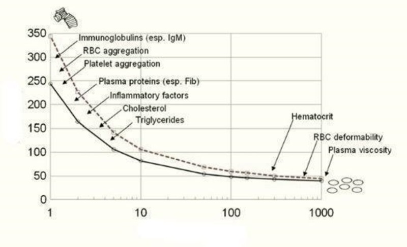
Blood viscosity is increased by elevated concentrations of acute phase reactants and hypergammaglobulinemia in inflammation. These increase blood viscosity by increasing plasma viscosity and fostering erythrocyte aggregation. Blood viscosity is also increased by decreased erythrocyte deformability, as occurs in malaria. Increased blood viscosity contributes to the association of acute infections with myocardial infarction (MI), venous thrombosis, and venous thromboembolism. It also increases vascular resistance, which decreases tissue perfusion and activates stretch receptors in the left ventricle, thereby initiating the systemic vascular resistance response. This compensates for the increased vascular resistance by vasodilation, lowering hematocrit, and decreasing intravascular volume. This physiological response causes the anemias associated with malaria, chronic inflammation, and other chronic diseases. Since tissue perfusion is inversely proportional to blood viscosity, anemia may be beneficial as it increases tissue perfusion when erythrocyte aggregating factors or erythrocytes with decreased deformability are present in the blood.
Keywords: acute phase response; anemia of chronic disease; atherosclerosis; atherothrombosis; blood viscosity; hemorheology; infection; inflammation; myocardial infarction; thrombosis.
Copyright © 2020, Sloop et al.
Conflict of interest statement
The authors have declared that no competing interests exist.
Figures
References
-
- Reducing cardiac after-load by lowering blood viscosity in patients with familial hypercholesterolemia - a pilot study. Possible mechanism for occurrence of anemia in chronic heart failure patients? Pop GAM, De Boer M-J, Stalenhoef AFH. Cor et Vasa. 2016;58:374–378.
-
- Sloop GD. Blood Viscosity: Its Role in Cardiovascular Pathophysiology and Hematology. Vol. 1. New York: Nova Science Publishers; 2017. The systemic vascular resistance response; pp. 67–82.
-
- Why atherothrombosis is in principle a hematologic disease: the effect of disorders and drugs which affect thrombosis on the development of atherosclerotic plaques. Sloop GD, Pop GAM, Weidman JJ, St Cyr JA. Int Arch Cardiovasc Dis. 2018;2
-
- Perspective: the failure of cholesteryl ester transfer protein inhibitors: is it due to increased blood viscosity? Sloop GD, Weidman JJ, St Cyr JA. Ther Adv Cardiovasc Dis. 2015;9:32–35. - PubMed
Publication types
LinkOut - more resources
Full Text Sources