Review
. 2020 Feb 24;12(2):e7092.
doi: 10.7759/cureus.7092.

A Review of the Theoretical Fascial Models: Biotensegrity, Fascintegrity, and Myofascial Chains

Affiliations
Review

A Review of the Theoretical Fascial Models: Biotensegrity, Fascintegrity, and Myofascial Chains

Bruno Bordoni et al. Cureus. .

Abstract

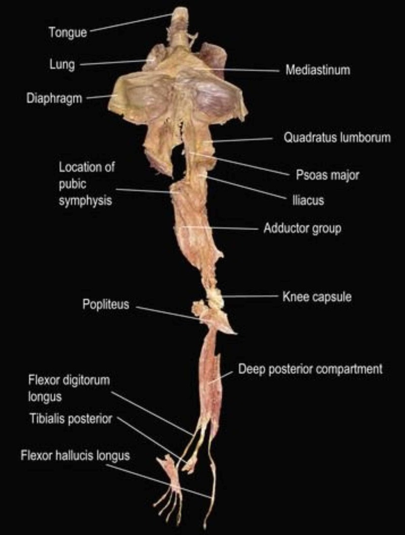
The fascial tissue includes solid and liquid fascia (body fluids such as blood and lymph). The fascia's nomenclature is the subject of debate in the academic world, as it is classified starting from different scientific perspectives. This disagreement is not a brake but is, in reality, the real wealth of research, the multidisciplinarity of thought and knowledge that leads to a deeper understanding of the topic. Another topic of discussion is the fascial model to conceptualize the human body, that is, how the fascial tissue fits into the living. Currently, there are some models: biotensegrity, fascintegrity, and myofascial chains. Biotensegrity is a mechanical model, which takes into consideration the solid fascia; fascintegrity considers the solid and the liquid fascia. Myofascial chains converge attention on the movement and transmission of force in the muscle continuum. The article is a reflection on fascial models and how these are theoretical-scientific visions that need to be further investigated.

Keywords: biotensegrety; fascia; fascintegrity; myofascial; myofascial chains; osteopathic; physiotherapy.


Conflict of interest statement

The authors have declared that no competing interests exist.

Figures

Figure 1
Figure 1. Shape and arrangement of the muscles on the ventral surface (a), dorsal (b) and lateral (c) of the human body
Reproduced with permission Anastasi et al. AA VV, Anatomia dell’uomo, fourth edition, 2010, pp76. Editor: Edi-Ermes, Milano [Human Anatomy].
Figure 2
Figure 2. Transverse section at the level of the upper third of the leg
Notes: 1 tibia; 2 muscular loggia; 3 interosseous membrane; 4 fibula; 5 intermuscular septum. All tissues are enveloped by fascial continuum. Reproduced with permission Anastasi et al. AA VV, Anatomia dell’uomo, fourth edition, 2010, pp. 89. Editor: Edi-Ermes, Milano [Human Anatomy].
Figure 3
Figure 3. Representation of the venous sinuses of the dura mater in a sagittal section of the skull (liquid fascia)
1 Vein of the dura mater; 7 Left superior petrosal sinus; 12 Straight sinus; 2 Superior sagittal sinus; 8 Left sigmoid sinus; 13 Great cerebral vein (of Galen); 3 Left middle cerebral vein; 9 Occipital sinus; 14 Inferior sagittal sinus; 4 Left sinus sphenoparietal; 10 Confluence of sinuses; 15 Superior sagittal sinus; 5 Intercavernous sinus; 11 Left transverse sinus; 16 Falx cerebri; 6 Left inferior petrosal sinus. Reproduced with permission Anastasi et al. AA VV, Anatomia dell’uomo, fourth edition, 2010, pp 432.  Editor: Edi-Ermes, Milano [Human Anatomy].
Figure 4
Figure 4. The figure shows the stretching of some parts of the body (the man in the picture is Dr Anthony Chrisco), and elongation is possible only if the whole body adapts to the district to be stretched (with the Fascianator method)
Photo was given by Dr Anthony Chrisco.
Figure 5
Figure 5. The figure shows how the whole body is held in tension to maintain the position, demonstrating the myofascial continuum (the woman in the picture is Dr Francesca Skyola)
The photo was given by Dr Francesca Skyola.
Figure 6
Figure 6. The figure illustrates the deep front line, which shows the continuous connection of myofascial tissue, from the muscles of the toes to the tongue
The image is taken from Thomas Myers' book: Anatomy Trains. Myofascial Meridians for Manual & Movement Therapists. Editor: Churchill Livingstone, 2013. Courtesy of Thomas Myers, co-author of the article.

References

    1. A case of fracture, attended with symptoms of unusual violence, relieved by an extensive longitudinal incision through the fascia of the limb. Mackesy J. https://www.ncbi.nlm.nih.gov/pubmed/30493501 Med Phys J. 1814;31:214–217. - PMC - PubMed
    1. New proposal to define the fascial system. Bordoni B, Marelli F, Morabito B, Castagna R, Sacconi B, Mazzucco P. Complement Med Res. 2018;25:257–262. - PubMed
    1. Fascial nomenclature:an update. Bordoni B, Walkowski S, Morabito B, Varacallo MA. Cureus. 2019;11:5718. - PMC - PubMed
    1. Improving the new definition of fascial system. Bordoni B. Complement Med Res. 2019;26:421–426. - PubMed
    1. A new concept of biotensegrity incorporating liquid tissues: blood and lymph. Bordoni B, Marelli F, Morabito B, Castagna R. J Evid Based Integr Med. 2018;23:2515690. - PMC - PubMed

LinkOut - more resources