. 2020 Feb 25;9(3):e393-e397.
doi: 10.1016/j.eats.2019.11.008. eCollection 2020 Mar.

The Outside-In, Percutaneous Release of the Medial Collateral Ligament for Knee Arthroscopy

Affiliations

The Outside-In, Percutaneous Release of the Medial Collateral Ligament for Knee Arthroscopy

Thomas E Moran et al. Arthrosc Tech. .

Abstract

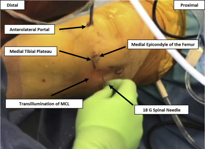
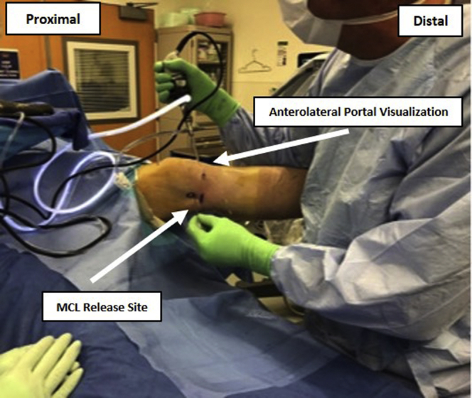
The outside-in, percutaneous release of the medial collateral ligament (MCL) is a technique used to increase the medial tibiofemoral joint space during arthroscopy to facilitate the use of instrumentation and improve visualization without causing iatrogenic cartilage damage. A recent systematic review of the literature has shown this technique to be efficacious and safe, with no evidence of associated short- or long-term complications. This technique has been used for this indication by the senior author without requiring any deviation from our institution's standard protocol for knee arthroscopy. In an attempt to standardize this technique's utilization and allow for further evaluation in the literature, the senior author's method for this percutaneous, outside-in approach of "pie crusting" the MCL is described.


Figures

Figure 1
Figure 1
An image of the medial left knee illustrates the landmarks used and the measurements made to identify the desired location for the percutaneous medial collateral ligament (MCL) release. The medial epicondyle of the femur and the medial tibial plateau are located by direct palpation and marked. The optimal location for the performance of the percutaneous release can then be measured relative to these landmarks with the use of a ruler.
Figure 2
Figure 2
Before performance of the percutaneous release, the arthroscope should be positioned in the anterolateral portal to permit adequate visualization of the medial compartment, while also providing transillumination of the medial joint space to further ensure accurate needle placement with respect to the marked landmarks and native knee anatomy. This setup is shown by an image before the percutaneous release of a right knee being performed. Abbreviation: MCL, medial collateral ligament.
Figure 3
Figure 3
After determining the optimal location for the performance of the medial collateral ligament (MCL) release, an 18-gauge spinal needle can be introduced superficially within 1 location, and translated subcutaneously, as indicated in the image of a right knee.
Figure 4
Figure 4
Subcutaneous fenestration is performed in an anterior to posterior direction to pie crust the medial collateral ligament (MCL), allowing for release. Depicted are the initial (left) and final (right) positions of the hand showing the lateral rotation of the wrist necessary for needle translation in the pie crust maneuver during release of a right knee MCL.
Figure 5
Figure 5
The medial joint space of the knee should be visualized simultaneously during the performance of this technique to ensure that an appropriate release of the medial collateral ligament (MCL) occurs, as illustrated in the image during a procedure on the left knee.
Figure 6
Figure 6
Intra-articular visualization through the anterolateral portal is used to confirm the adequacy of the release performed on the medial collateral ligament (MCL) and can be used to ensure appropriate access to the injury of interest. This image provides an intra-articular view of the medial compartment width before (left) and width generated after performance of the MCL release (right) on the right knee.

References

    1. Mordecai S.C., Al-Hadithy N., Ware H.E., Gupte C.M. Treatment of meniscal tears: An evidence based approach. World J Orthop. 2014;5:233–241. - PMC - PubMed
    1. Moran T.E., Awowale J.T., Werner B.C., Fox M.A., Miller M.D. Associated morbidity after the percutaneous release of the medial collateral ligament for knee arthroscopy. Arthrosc. 2020;36:891–900. - PubMed
    1. Polat B., Deniz A., Polat A.E. Objective measurement of medial joint space widening with percutaneous “pie crust” release of medial collateral ligament during knee arthroscopy. J Knee Surg. 2020;33:94–98. - PubMed
    1. Ward B.D., Lubowitz J.H. Basic knee arthroscopy part 3: Diagnostic arthroscopy. Arthrosc Tech. 2013;2:e503–e505. - PMC - PubMed
    1. LaPrade R.F., Engebretsen A.H., Ly T.V., Johansen S., Wentorf F.A., Engebretsen L. The anatomy of the medial part of the knee. J Bone Jt Surg Am. 2007;89:2000–2010. - PubMed