. 2020 Jun;17(3):323-333.
doi: 10.1007/s13770-020-00244-w. Epub 2020 Mar 29.

Engineered M13 Peptide Carrier Promotes Angiogenic Potential of Patient-Derived Human Cardiac Progenitor Cells and In Vivo Engraftment

Affiliations

Engineered M13 Peptide Carrier Promotes Angiogenic Potential of Patient-Derived Human Cardiac Progenitor Cells and In Vivo Engraftment

Woong Bi Jang et al. Tissue Eng Regen Med. 2020 Jun.

Abstract

Background: Despite promising advances in stem cell-based therapy, the treatment of ischemic cardiovascular diseases remains a big challenge due to both the insufficient in vivo viability of transplanted cells and poor angiogenic potential of stem cells. The goal of this study was to develop therapeutic human cardiac progenitor cells (hCPCs) for ischemic cardiovascular diseases with a novel M13 peptide carrier.

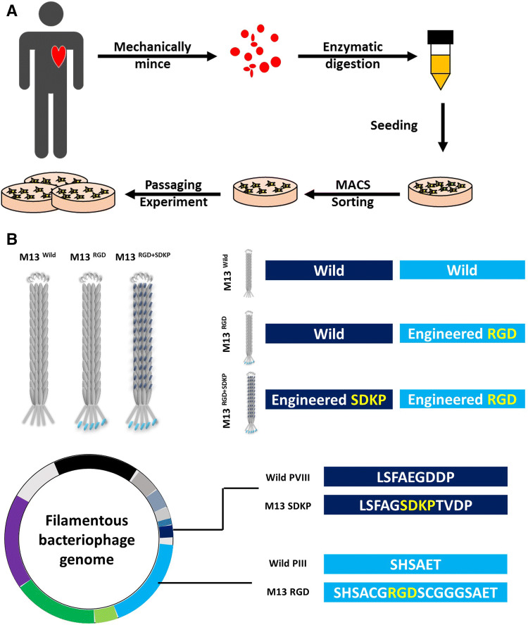
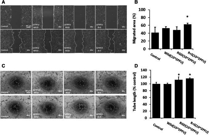
Method: In this study, an engineered M13 peptide carrier was successfully generated using a QuikChange Kit. The cellular function of M13 peptide carrier-treated hCPCs was assessed using a tube formation assay and scratch wound healing assay. The in vivo engraftment and cell survival bioactivities of transplanted cells were demonstrated by immunohistochemistry after hCPC transplantation into a myocardial infarction animal model.

Results: The engineered M13RGD+SDKP peptide carrier, which expressed RGD peptide on PIII site and SDKP peptide on PVIII site, did not affect morphologic change and proliferation ability in hCPCs. In contrast, hCPCs treated with M13RGD+SDKP showed enhanced angiogenic capacity, including tube formation and migration capacity. Moreover, transplanted hCPCs with M13RGD+SDKP were engrafted into the ischemic region and promoted in vivo cell survival.

Conclusion: Our present data provides a promising protocol for CPC-based cell therapy via short-term cell priming of hCPCs with engineered M13RGD+SDKP before cell transplantation for treatment of cardiovascular disease.

Keywords: Cardiac progenitor cell; Cardiovascular diseases; M13 bacteriophage; RGD; SDKP.


Conflict of interest statement

The authors have no financial conflicts of interest.

Figures

Fig. 1
Fig. 1
Experimental scheme of this study. A Isolation protocol of primary human cardiac progenitor cells. Biopsied heart specimens were minced mechanically into small pieces, digested with collagenase, and sorted by MACS. B Design and characterization of engineered m13 phage. The M13 phage was engineered to express RGD-peptide on PIII site and SDKP on PVIII site
Fig. 2
Fig. 2
Effects of engineered M13 treatment on human CPCs. A hCPCs were treated with different concentrations of M13 nano-carriers for 24 h and viability was measured using a cell viability assay (CCK assay). Data are presented as mean ± standard deviation (SD). *, p < 0.05 versus control. B Expression of M13 nano-carrier in hCPCs pretreated with M13Wild, RGD and RGD+SDKP by immunocytochemistry. Scale bar = 20 µm. C Quantification of adhesion of the engineered M13 phage at various concentrations (0–106 plaque forming unit) on the CPCs. D hCPCs were treated with M13Wild, RGD and RGD+SDKP for 24 h and morphologic changes were confirmed using a microscope. Scale bar = 50 µm. E Quantification of the diameter of the hCPCs treated with the engineered M13 nano-carrier
Fig. 3
Fig. 3
Effect of engineered M13 nano-carrier on migration and angiogenic capacity of hCPCs. A The migration capacity of the hCPCs treated with the engineered M13 was investigated using a scratched wound healing assay. Scale bar = 50 µm. B Quantification of migrated area. Data are presented as mean ± standard deviation (SD). *, p < 0.05 versus control. C The tube formation capacity of the hCPCs treated with the engineered M13 nano-carrier was assessed using a Matrigel tube formation assay. Scale bar = 50 µm. D Quantification of total tube length of the hCPCs treated with the engineered M13 nano-carrier
Fig. 4
Fig. 4
Engraftment of human CPCs RGD + SDKP enhanced heart function recovery after 28 days post-MI. A At 28 days after MI, heart tissue was harvested. Heart sections were stained with Masson’s trichrome dye. Representative whole-heart images for serial sections. Scale bar = 2000 μm. B SMA-positive blood vessels in ischemic samples on day 28. Scale bar = 100 μm. C Quantitative analysis of fibrosis and SMA-positive blood vessels. Average values presented as mean ± SD (n = 5). *p < 0.05, **p < 0.01
Fig. 5
Fig. 5
Analysis of survival and proliferation of implanted human CPCs RGD + SDKP. A Representative DiI + CPCs (orange) after injection with M13 (WT, RGD, RGD + SDKP) at 3 days post-infarction. Phosphorylated histone H3 (pHH3; green), sarcomeric alpha-sctin (α-SA; red) and nuclei were stained using DAPI. B, C The statistics of DiI + MNC retention ratio and quantification of pHH3-positive cells at 3 days after MI. Data are presented as mean ± SD (n = 5). *p < 0.05, **p < 0.01. Scale bar = 100 μm
Fig. 6
Fig. 6
Engraftment of human CPCs RGD + SDKP enhanced neovascularization 3 days post-MI. A Representative images of ischemic heart samples at 3 days after immunostaining with anti-CD31 antibody (red) and anti-SMA antibody (green). Nuclei counterstained with DAPI (blue). B, C Quantitative analysis of CD31-positive capillaries. B SMA-positive blood vessels (C) in ischemic samples on day 3. Average values presented as mean ± SD (n = 5). *p < 0.05, **p < 0.01. Scale bar = 100 μm

References

    1. Bhatnagar A. Environmental determinants of cardiovascular disease. Circ Res. 2017;121:162–180. - PMC - PubMed
    1. Cohen BE, Edmondson D, Kronish IM. State of the art review: depression, stress, anxiety, and cardiovascular disease. Am J Hypertens. 2015;28:1295–1302. - PMC - PubMed
    1. Moran AE, Roth GA, Narula J, Mensah GA. 1990–2010 Global cardiovascular disease Atlas. Glob Heart. 2014;9:3–16. - PubMed
    1. Thomas H, Diamond J, Vieco A, Chaudhuri S, Shinnar E, Cromer S, et al. Global Atlas of cardiovascular disease 2000–2016: the path to prevention and control. Glob Heart. 2018;13:143–163. - PubMed
    1. De Caterina R, Zampolli A, Del Turco S, Madonna R, Massaro M. Nutritional mechanisms that influence cardiovascular disease. Am J Clin Nutr. 2006;83:421S–426S. - PubMed

Publication types

LinkOut - more resources