

Immunization with virus-like particles conjugated to CIDRα1 domain of Plasmodium falciparum erythrocyte membrane protein 1 induces inhibitory antibodies
- PMID: 32228596
- PMCID: PMC7106694
- DOI: 10.1186/s12936-020-03201-z
Immunization with virus-like particles conjugated to CIDRα1 domain of Plasmodium falciparum erythrocyte membrane protein 1 induces inhibitory antibodies
Abstract
Background: During the erythrocytic cycle, Plasmodium falciparum malaria parasites express P. falciparum Erythrocyte Membrane Protein 1 (PfEMP1) that anchor the infected erythrocytes (IE) to the vascular lining of the host. The CIDRα1 domain of PfEMP1 is responsible for binding host endothelial protein C receptor (EPCR), and increasing evidence support that this interaction triggers severe malaria, accounting for the majority of malaria-related deaths. In high transmission regions, children develop immunity to severe malaria after the first few infections. This immunity is believed to be mediated by antibodies targeting and inhibiting PfEMP1, causing infected erythrocytes to circulate and be cleared in the spleen. The development of immunity to malaria coincides with acquisition of broad antibody reactivity across the CIDRα1 protein family. Altogether, this identifies CIDRα1 as an important vaccine target. However, the antigenic diversity of the CIDRα1 domain family is a challenge for vaccine development.
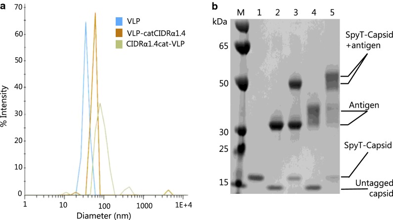
Methods: Immune responses in mice vaccinated with Virus-Like Particles (VLP) presenting CIDRα1 antigens were investigated. Antibody reactivity was tested to a panel of recombinant CIDRα1 domains, and the antibodies ability to inhibit EPCR binding by the recombinant CIDRα1 domains was tested in Luminex-based multiplex assays.
Results: VLP-presented CIDRα1.4 antigens induced a rapid and strong IgG response capable of inhibiting EPCR-binding of multiple CIDRα1 domains mainly within the group A CIDRα1.4-7 subgroups.
Conclusions: The study observations mirror those from previous CIDRα1 vaccine studies using other vaccine constructs and platforms. This suggests that broad CIDRα1 antibody reactivity may be achieved through vaccination with a limited number of CIDRα1 variants. In addition, this study suggest that this may be achieved through vaccination with a human compatible VLP vaccine platform.
Keywords: Antigenic diversity; CIDRα1; Malaria; PfEMP1; Plasmodium falciparum; Vaccine; Virus-like particle.
Conflict of interest statement
The authors have no competing interests.
Figures


Similar articles
-
Mosaic and cocktail capsid-virus-like particle vaccines for induction of antibodies against the EPCR-binding CIDRα1 domain of PfEMP1.PLoS One. 2024 Jul 24;19(7):e0302243. doi: 10.1371/journal.pone.0302243. eCollection 2024. PLoS One. 2024. PMID: 39046960 Free PMC article.
-
Immunization with Recombinant Plasmodium falciparum Erythrocyte Membrane Protein 1 CIDRα1 Domains Induces Domain Subtype Inhibitory Antibodies.Infect Immun. 2018 Oct 25;86(11):e00435-18. doi: 10.1128/IAI.00435-18. Print 2018 Nov. Infect Immun. 2018. PMID: 30150256 Free PMC article.
-
Identification of broadly inhibitory anti-PfEMP1 antibodies by mass spectrometry sequencing of plasma IgG from a malaria-exposed child.Proc Natl Acad Sci U S A. 2025 Aug 26;122(34):e2508744122. doi: 10.1073/pnas.2508744122. Epub 2025 Aug 20. Proc Natl Acad Sci U S A. 2025. PMID: 40833410
-
Broadly inhibitory antibodies to severe malaria virulence proteins.Nature. 2024 Dec;636(8041):182-189. doi: 10.1038/s41586-024-08220-3. Epub 2024 Nov 20. Nature. 2024. PMID: 39567685 Free PMC article.
-
PfEMP1 - A Parasite Protein Family of Key Importance in Plasmodium falciparum Malaria Immunity and Pathogenesis.Adv Parasitol. 2015 Apr;88:51-84. doi: 10.1016/bs.apar.2015.02.004. Epub 2015 Mar 23. Adv Parasitol. 2015. PMID: 25911365 Review.
Cited by
-
Evasive mechanisms of human VSG and PfEMP1 antigens with link to Vaccine scenario: a review.J Parasit Dis. 2025 Mar;49(1):13-28. doi: 10.1007/s12639-024-01740-9. Epub 2024 Sep 24. J Parasit Dis. 2025. PMID: 39975623 Review.
-
Virus-like particles expressing microneme-associated antigen of Plasmodium berghei confer better protection than those expressing apical membrane antigen 1.Parasites Hosts Dis. 2024 May;62(2):193-204. doi: 10.3347/PHD.24017. Epub 2024 May 27. Parasites Hosts Dis. 2024. PMID: 38835260 Free PMC article.
-
Malaria: past, present, and future.Signal Transduct Target Ther. 2025 Jun 17;10(1):188. doi: 10.1038/s41392-025-02246-3. Signal Transduct Target Ther. 2025. PMID: 40523953 Free PMC article. Review.
-
Mosaic and cocktail capsid-virus-like particle vaccines for induction of antibodies against the EPCR-binding CIDRα1 domain of PfEMP1.PLoS One. 2024 Jul 24;19(7):e0302243. doi: 10.1371/journal.pone.0302243. eCollection 2024. PLoS One. 2024. PMID: 39046960 Free PMC article.
-
Structure-Guided Design of a Synthetic Mimic of an Endothelial Protein C Receptor-Binding PfEMP1 Protein.mSphere. 2021 Jan 6;6(1):e01081-20. doi: 10.1128/mSphere.01081-20. mSphere. 2021. PMID: 33408232 Free PMC article.
References
-
- Bruce-Chwatt LJ. A longitudinal survey of natural malaria infection in a group of West African adults. West Afr Med J. 1963;12:199–217. - PubMed
MeSH terms
Substances
Grants and funding
LinkOut - more resources
Full Text Sources
Other Literature Sources
Medical
Research Materials
Miscellaneous
