

Case Reports
doi: 10.4269/ajtmh.19-0861.
Case Report: Mucosal Leishmaniasis in New York City
Affiliations
- PMID: 32228792
- PMCID: PMC7253091
- DOI: 10.4269/ajtmh.19-0861
Item in Clipboard
Case Reports
Case Report: Mucosal Leishmaniasis in New York City
Am J Trop Med Hyg.
2020 Jun.
Abstract
The six previously reported civilian cases of mucosal leishmaniasis (ML) diagnosed in the United States have all represented imported New World ML. We describe two new patients with ML diagnosed in New York City-a Syrian immigrant with a nasal mass (Leishmania tropica), the first report of Old World ML in the United States, and an American ecologist who worked in Bolivia and had been treated for cutaneous infection 23 years before developing lesions (L. (Viannia) braziliensis) initially of the uvula, soft palate, and posterior pharynx and subsequently the larynx.
Figures
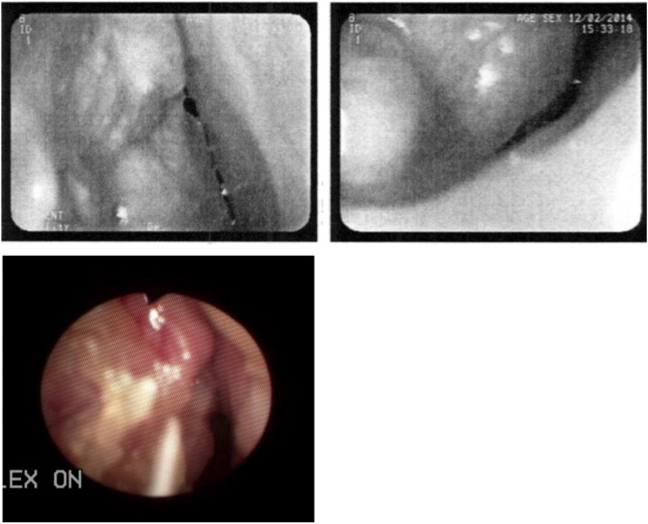
Patient 1. Pretreatment endoscopic views of lobulated left nasal mass obtained before (top) and after referral to Weill Cornell Medical College (bottom, left). This figure appears in color at www.ajtmh.org .

Photomicrographs of hematoxylin and eosin–stained tissue sections from biopsy of nasal mass. Top: Note mononuclear cell inflammatory infiltrate and numerous intracellular amastigotes, particularly within enlarged macrophage phagocytic vacuoles (arrows). Bottom: High-power view showing intracellular amastigotes within phagocytic vacuoles; arrow indicates an amastigote showing characteristic oval shape and darkly stained round nucleus and rod-shaped kinetoplast. Original magnifications: ×400 (top) and ×630 (bottom). This figure appears in color at www.ajtmh.org .

Patient 2. Left: Postoperative photograph showing absent uvula and ulcerated and cobblestone appearance of soft palate and posterior pharyngeal wall. Preoperatively (photographs not performed), the uvula showed similar changes. Right: Laryngoscopic view 11 months after initial treatment showing edematous right aryepiglottic fold and mucosal cobblestoning (circled) of the laryngeal ventricle. This figure appears in color at www.ajtmh.org .
References
-
- Singer C, Armstrong D, Jones TC, Spiro RH, 1975. Imported mucocutaneous leishmaniasis in New York city. Report of a patient treated with amphotericin B. Am J Med 59: 444–447. - PubMed
-
- Huna-Baron R, Warren FA, Miller W, Jacobs J, Green J, Kupersmith MJ, 2000. Mucosal leishmaniasis presenting as sinusitis and optic neuropathy. Arch Ophthalmol 118: 852–854. - PubMed
-
- Costa JW, Milner DA, Maguire JH, 2003. Mucocutaneous leishmaniasis in a US citizen. Oral Surg Oral Med Oral Pathol Oral Radiol Endod 96: 573–577. - PubMed
-
- Brahn E, Pegues DA, Craft N, 2010. Mucocutaneous leishmaniasis as Wegener’s granulomatosis. J Clin Rheumatol 16: 125–128. - PubMed
-
- Stein DJ, Yarlagadda BB, Noordzij JP, 2013. Otolaryngologic presentation of mucosal leishmaniasis. J Case Rep Med 1: 1–2.
Publication types
MeSH terms
Substances
LinkOut - more resources
Full Text Sources
