. 2020 Mar 31:9:e54639.
doi: 10.7554/eLife.54639.

Approaching boiling point stability of an alcohol dehydrogenase through computationally-guided enzyme engineering

Affiliations

Approaching boiling point stability of an alcohol dehydrogenase through computationally-guided enzyme engineering

Friso S Aalbers et al. Elife. .

Abstract

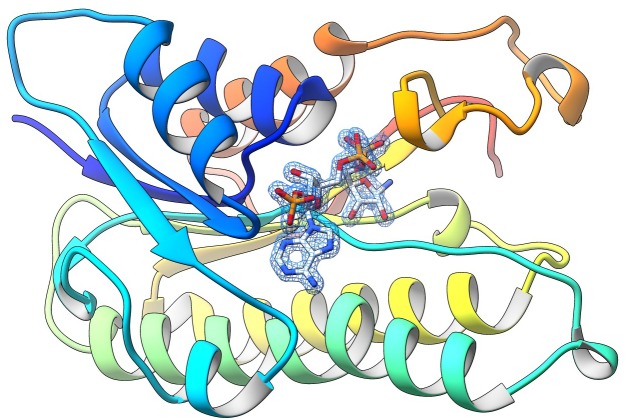
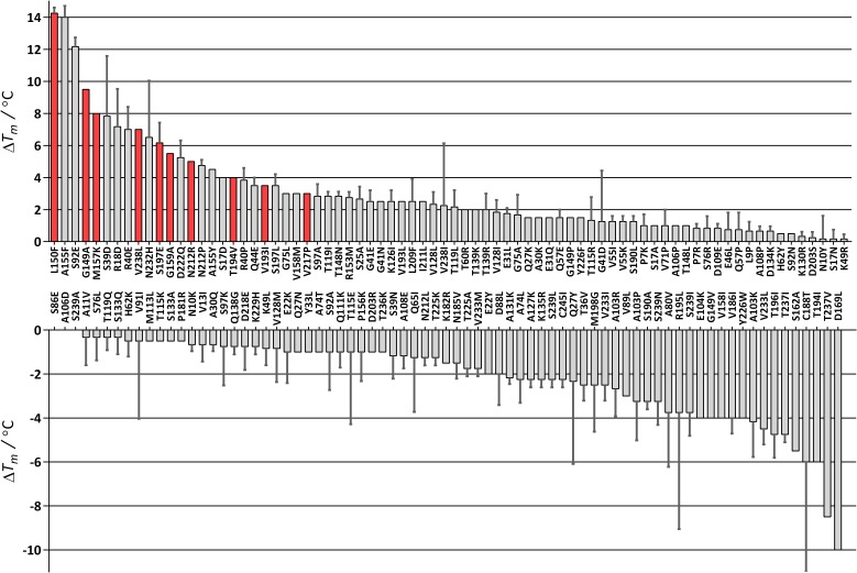
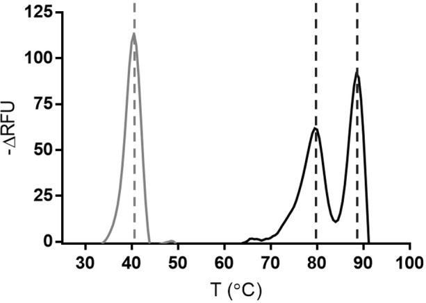
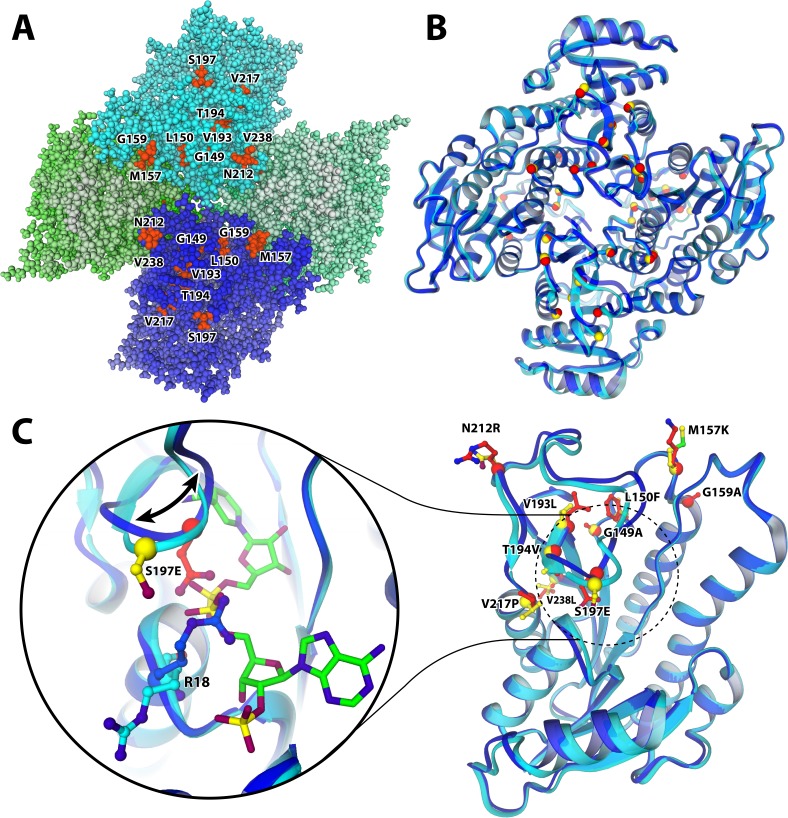
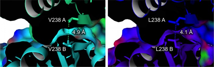
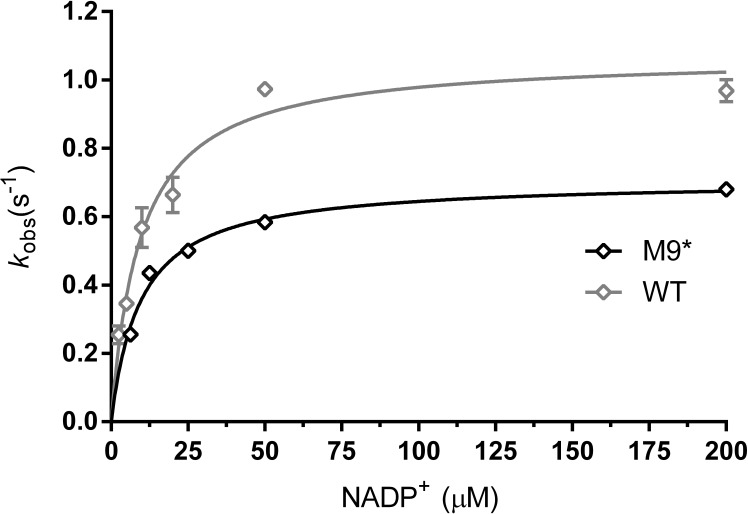
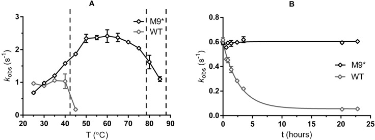
Enzyme instability is an important limitation for the investigation and application of enzymes. Therefore, methods to rapidly and effectively improve enzyme stability are highly appealing. In this study we applied a computational method (FRESCO) to guide the engineering of an alcohol dehydrogenase. Of the 177 selected mutations, 25 mutations brought about a significant increase in apparent melting temperature (ΔTm ≥ +3 °C). By combining mutations, a 10-fold mutant was generated with a Tm of 94 °C (+51 °C relative to wild type), almost reaching water's boiling point, and the highest increase with FRESCO to date. The 10-fold mutant's structure was elucidated, which enabled the identification of an activity-impairing mutation. After reverting this mutation, the enzyme showed no loss in activity compared to wild type, while displaying a Tm of 88 °C (+45 °C relative to wild type). This work demonstrates the value of enzyme stabilization through computational library design.

Keywords: E. coli; alcohol dehydrogenase; biocatalysis; biotechnology; cofactor; computational biology; enzyme engineering; molecular biophysics; oxidations; structural biology; systems biology.


Conflict of interest statement

FA, MF, SR, MT, JG, AV, AM, MF No competing interests declared, SB A patent application on the original ADH was filed by c-LEcta (WO 2019/012095)

Figures

Figure 1.
Figure 1.. The crystal structure of ADHA.
The figure highlights the final weighted 2Fo-Fc map for NADP+ bound to a subunit of the wild-type ADHA (subunit A, contour level 1.2 σ). The nicotinamide moiety of the cofactor is disordered and was not included in the final model.
Figure 2.
Figure 2.. Difference in Tm for 151 FRESCO-predicted ADHA mutants.
The average of two measurements is given and the standard error. The Tm of wild-type ADHA is 43 °C (set as 0). The 10 stabilizing mutations with a red bar were combined. Melting curves of wild type and the final mutant (M9*) are depicted in Figure 2—figure supplement 1.
Figure 2—figure supplement 1.
Figure 2—figure supplement 1.. Melting curves.
Apparent melting temperatures of wild type (in grey) and M9* variant. RFU = relative fluorescence units. M9* displays a first melting peak at 79 °C, second peak at 88 °C. One curve is shown from three technical replicates.
Figure 3.
Figure 3.. Michaelis-Menten plots for kinetics with NADP+.
(A) ADHA wild type (B) M9 mutant. Note that the X-axis scaling is different. The inset of B presents the same data with a different Y-axis scaling. Plots are fitted with Michaelis-Menten in GraphPad prism 6.07.
Figure 4.
Figure 4.. Structure of the M9 mutant of ADHA with mutated resides highlighted.
(A and B) quaternary structure of M9. The tetramer is organized such that the N-termini are on the outside (on the edge of the top-down view of A and B), whereas the C-termini all point inwards; which is where most and the most stabilizing mutations were found. (A) M9 structure with all atoms represented as balls. The four monomers are shaded in various colours, highlighting the particular clustering of the observed stabilizing mutations. (B) The structure as ribbon model, superimposing the mutant (blue ribbon, red spheres indicate mutated residue) and the wild type (cyan ribbon, yellow spheres). (C) Colour scheme as in B. The loop (196-214) that is dislocated as a result of the S197E mutation, compared to the structure of wild-type ADHA. The shift is accompanied by a flip of R18 into the NADP-binding pocket, likely due to an electrostatic attraction from the mutant glutamate. As a result, the cofactor (green carbons) is only bound in the wild type, while absent from the mutant structure.
Figure 5.
Figure 5.. Dimer interface with the V238L mutation (ΔTm = 7 ˚C).
(A and B) indicate the different monomers in the ADHA tetramer.
Figure 6.
Figure 6.. Michaelis-Menten plots for kinetics with NADP+.
ADHA wild type (grey, triangles) and M9* mutant (M9 with S197E reverted) (black, diamonds). Plots are fitted with the Michaelis-Menten equation in GraphPad prism 6.07.
Figure 7.
Figure 7.. Properties of wild-type and M9* ADHA.
(A) Temperature-activity profile using cyclohexanol as substrate. The dashed lines indicate the Tof the wild type (at 43 °C) and apparent melting temperatures of M9* (78.5 °C and 88 °C) (B) Enzyme activity monitored over time at 37 °C (buffer composition: 50 mM Tris-HCl pH 7.5). Figure 7—figure supplement 1 depicts the enzyme activity over time for 18 days.
Figure 7—figure supplement 1.
Figure 7—figure supplement 1.. Long-term stability.
Long-term stability at 37 °C of the wild-type protein (in grey, triangles) compared to M9* (in black, diamonds). Plot fitted with one-phase decay in GraphPad prism 6.07.

References

    1. Arabnejad H, Lago MD, Jekel PA, Floor RJ, Thunnissen A, Terwisscha Van Scheltinga AC, Wijma HJ, Janssen DB. A robust cosolvent-compatible halohydrin dehalogenase by computational library design. Protein Engineering, Design & Selection : PEDS. 2017;30:175–189. doi: 10.1093/protein/gzw068. - DOI - PubMed
    1. Bednar D, Beerens K, Sebestova E, Bendl J, Khare S, Chaloupkova R, Prokop Z, Brezovsky J, Baker D, Damborsky J. FireProt: energy- and Evolution-Based computational design of thermostable Multiple-Point mutants. PLOS Computational Biology. 2015;11:e1004556. doi: 10.1371/journal.pcbi.1004556. - DOI - PMC - PubMed
    1. Bhatia C, Oerum S, Bray J, Kavanagh KL, Shafqat N, Yue W, Oppermann U. Towards a systematic analysis of human short-chain dehydrogenases/reductases (SDR): Ligand identification and structure-activity relationships. Chemico-Biological Interactions. 2015;234:114–125. doi: 10.1016/j.cbi.2014.12.013. - DOI - PubMed
    1. Bjørk A, Dalhus B, Mantzilas D, Sirevåg R, Eijsink VG. Large improvement in the thermal stability of a tetrameric malate dehydrogenase by single point mutations at the dimer-dimer interface. Journal of Molecular Biology. 2004;341:1215–1226. doi: 10.1016/j.jmb.2004.06.079. - DOI - PubMed
    1. Bommarius AS, Paye MF. Stabilizing biocatalysts. Chemical Society Reviews. 2013;42:6534–6565. doi: 10.1039/c3cs60137d. - DOI - PubMed

Publication types

Substances

Supplementary concepts

Grants and funding