

Diagnosis of the Coronavirus disease (COVID-19): rRT-PCR or CT?
- PMID: 32229322
- PMCID: PMC7102545
- DOI: 10.1016/j.ejrad.2020.108961
Diagnosis of the Coronavirus disease (COVID-19): rRT-PCR or CT?
Abstract
Purpose: To evaluate the diagnostic value of computed tomography (CT) and real-time reverse-transcriptase-polymerase chain reaction (rRT-PCR) for COVID-19 pneumonia.
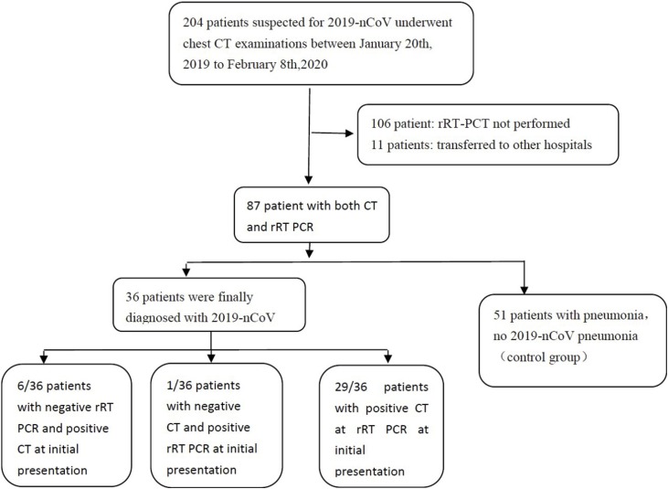
Methods: This retrospective study included all patients with COVID-19 pneumonia suspicion, who were examined by both CT and rRT-PCR at initial presentation. The sensitivities of both tests were then compared. For patients with a final confirmed diagnosis, clinical and laboratory data, in addition to CT imaging findings were evaluated.
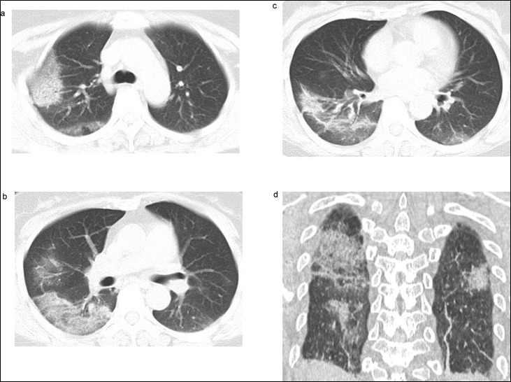
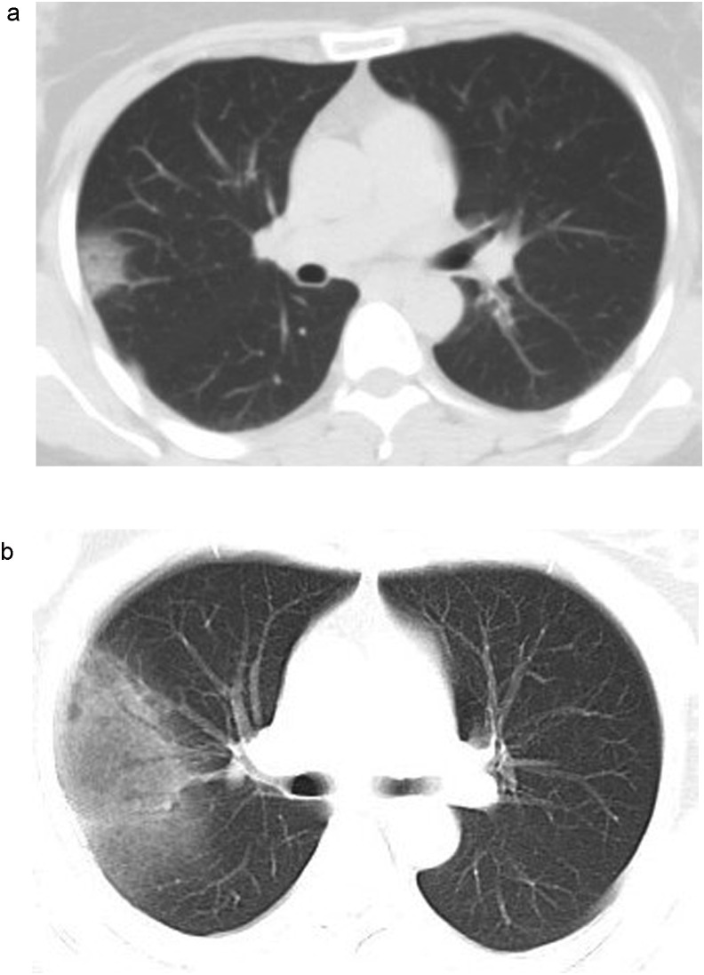
Results: A total of 36 patients were finally diagnosed with COVID-19 pneumonia. Thirty-five patients had abnormal CT findings at presentation, whereas one patient had a normal CT. Using rRT-PCR, 30 patients were tested positive, with 6 cases initially missed. Amongst these 6 patients, 3 became positive in the second rRT-PCR assay(after 2 days, 2 days and 3 days respectively), and the other 3 became positive only in the third round of rRT-PCR tests(after 5 days, 6 days and 8 days respectively). At presentation, CT sensitivity was therefore 97.2%, whereas the sensitivity of initial rRT-PCR was only 83.3%.
Conclusion: rRT-PCR may produce initial false negative results. We suggest that patients with typical CT findings but negative rRT-PCR results should be isolated, and rRT-PCR should be repeated to avoid misdiagnosis.
Keywords: Coronavirus; Pneumonia; Severe Acute Respiratory Syndrome; Tomography; X-Ray Computed.
Copyright © 2020 Elsevier B.V. All rights reserved.
Figures
References
-
- Rubin E.J., Baden L.R., Morrissey S., Campion E.W. Medical Journals and the 2019-nCoV Outbreak. N Engl J Med. 2020 - PubMed
MeSH terms
Substances
LinkOut - more resources
Full Text Sources
Other Literature Sources
