. 2020 May 1;295(18):6214-6224.
doi: 10.1074/jbc.RA120.013067. Epub 2020 Mar 30.

A Legionella effector kinase is activated by host inositol hexakisphosphate

Affiliations

A Legionella effector kinase is activated by host inositol hexakisphosphate

Anju Sreelatha et al. J Biol Chem. .

Abstract

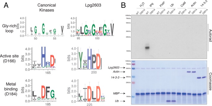
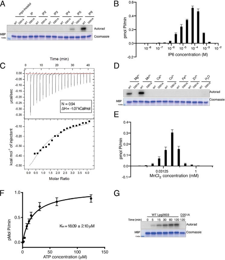
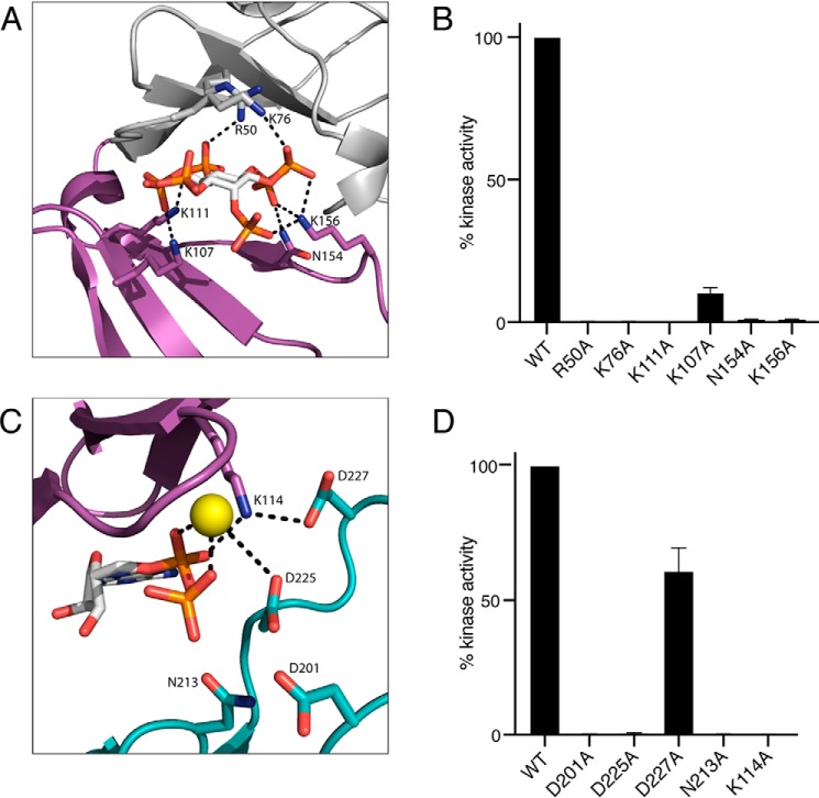
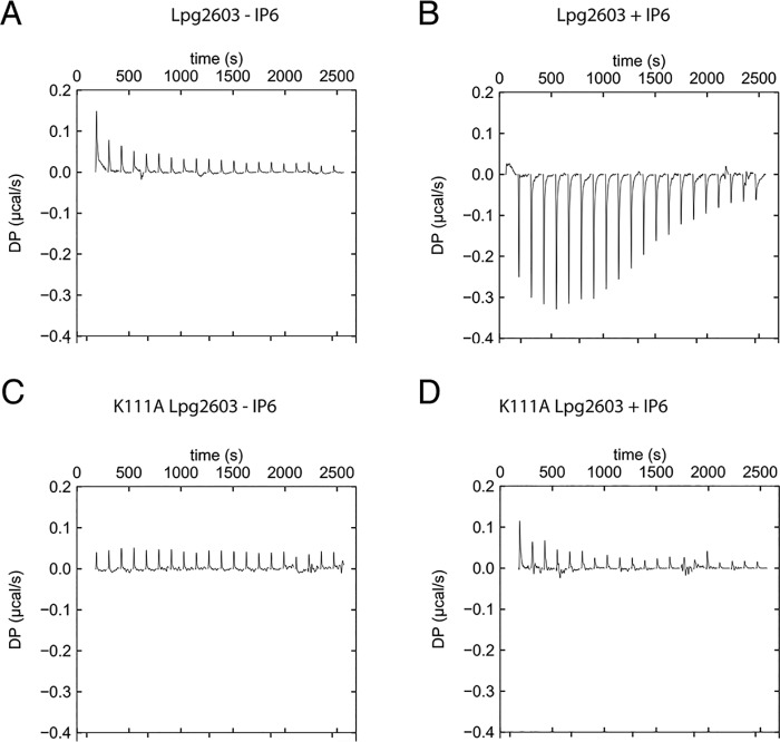
The transfer of a phosphate from ATP to a protein substrate, a modification known as protein phosphorylation, is catalyzed by protein kinases. Protein kinases play a crucial role in virtually every cellular activity. Recent studies of atypical protein kinases have highlighted the structural similarity of the kinase superfamily despite notable differences in primary amino acid sequence. Here, using a bioinformatics screen, we searched for putative protein kinases in the intracellular bacterial pathogen Legionella pneumophila and identified the type 4 secretion system effector Lpg2603 as a remote member of the protein kinase superfamily. Employing an array of biochemical and structural biology approaches, including in vitro kinase assays and isothermal titration calorimetry, we show that Lpg2603 is an active protein kinase with several atypical structural features. Importantly, we found that the eukaryote-specific host signaling molecule inositol hexakisphosphate (IP6) is required for Lpg2603 kinase activity. Crystal structures of Lpg2603 in the apo-form and when bound to IP6 revealed an active-site rearrangement that allows for ATP binding and catalysis. Our results on the structure and activity of Lpg2603 reveal a unique mode of regulation of a protein kinase, provide the first example of a bacterial kinase that requires IP6 for its activation, and may aid future work on the function of this effector during Legionella pathogenesis.

Keywords: Legionella; allosteric regulation; bacterial pathogenesis; bacterial protein kinase; effector; enzyme structure; inositol hexikisphosphate; inositol phosphate.


Conflict of interest statement

The authors declare that they have no conflicts of interest with the contents of this article

Figures

Figure 1.
Figure 1.
Lpg2603 is an active kinase in the presence of IP6. A, sequence logo built using 74 homologs of Lpg2603 depicting conserved motifs present in kinases including the Gly-rich loop and active site residues (numbering in parentheses corresponds to PKA). The logo for canonical kinases was built using 3998 homologs (Pfam domain PF00069). B, autoradiograph depicting the incorporation of 32P from [γ-32P]ATP into MBP by recombinant Lpg2603 WT or the inactive mutant, D201A, in the presence of different cofactors: IP6, PI4P, ubiquitin (Ub), calmodulin (CaM), globular actin, or 14-3-3 protein. Note that CaM migrates at the same molecular weight as MBP. The reactions were terminated after 10 min by the addition of EDTA, and the products were resolved by SDS-PAGE and visualized by Coomassie Blue staining (lower panel) and autoradiography (upper panel). The results are representative of three independent experiments.
Figure 2.
Figure 2.
Lpg2603 activity requires IP6 and Mn2+. A, in vitro kinase assay showing incorporation of 32P from [γ-32P]ATP into MBP by Lpg2603 in the presence of myo-inositol, inositol phosphate (IP), inositol diphosphate (IP2), inositol triphosphate (IP3), inositol tetraphosphate (IP4), inositol pentaphosphate (IP5), and IP6. The reactions were terminated and analyzed as in Fig. 1B. The results are representative of three independent experiments. B, incorporation of γ-32P from [γ-32P]ATP into MBP by Lpg2603 with increasing concentrations of IP6. The reactions were terminated after 10 min by the addition of EDTA, the products were resolved by SDS-PAGE, and Coomassie-stained MBP bands were excised for scintillation counting. The results are representative of three independent experiments. The error bars represent standard deviation from three replicates from an individual experiment. C, representative isothermal calorimetry data for Lpg2603 binding to IP6. Lpg2603 is at 200 μm in the cell, and IP6 is at 8 mm in the titration syringe for a final molar ratio of 1:4. Best fit parameters were as follows: n = 0.94; ΔH = −1.07 kCal/mol. D, in vitro kinase assay showing incorporation of γ-32P from [γ-32P]ATP into MBP by Lpg2603 in the presence or absence of MgCl2, MnCl2, CaCl2, CoCl2, FeCl2, and ZnCl2. The reactions were terminated and analyzed as in Fig. 1B. The results are representative of three independent experiments. E, incorporation of γ-32P from [γ-32P]ATP into MBP by Lpg2603 with varying concentrations of MnCl2. The reactions were terminated and analyzed as in B. The results are representative of three independent experiments. The error bars represent standard deviation from three replicates from an individual experiment. F, kinetic analysis depicting the concentration dependence of Mn2+/ATP on the rate of MBP phosphorylation by Lpg2603. Km for Mn2+/ATP = 18.09 ± 2.10 μm; Vmax = 1.05 ± 0.04 pmol/min Reactions were terminated and analyzed as in B. The results are representative of three independent experiments. The error bars represent standard error of the mean from three replicates from an individual experiment. Kinetic measurements were fitted into the Michaelis–Menten equation using GraphPad Prism. G, time-dependent incorporation of γ-32P from [γ-32P]ATP into MBP by Lpg2603. The reactions were terminated and analyzed as in Fig. 1B. The results are representative of three independent experiments.
Figure 3.
Figure 3.
Crystal structure of Lpg2603 reveals a unique mode of kinase activation by IP6. A, ribbon representation of apo-Lpg2603. The N-lobe and C-lobe are depicted in magenta and teal, respectively. The αC-helix and the N-terminal extensions are shown in orange and white, respectively. B, ribbon representation of Lpg2603 bound to IP6. The ligand IP6 is shown in ball-and-stick form, colored according to atom. C, ribbon representation of Lpg2603 bound to IP6 and ADP. The nucleotide ADP and ligand IP6 are shown in ball-and-stick form, colored according to atom. The Mn2+ ion is shown as a yellow sphere. D, superposition of the apo-Lpg2603 (shown in pink) with the nucleotide and ligand-bound Lpg2603 (shown in green) depicting the disordered N-lobe in the apo structure.
Figure 4.
Figure 4.
Mutational analysis highlights residues in Lpg2603 important for catalysis. A, enlarged image of IP6-binding pocket showing residues and interactions important for IP6 binding. IP6 is shown in ball-and-stick form, colored according to atom. B, activity of Lpg2603 or mutants were assayed as in Fig. 2B. Activity is expressed relative to the WT enzyme. The results are representative of three independent experiments. The error bars represent standard deviation. C, enlarged image of the nucleotide-binding pocket showing residues and interactions important for ADP binding and catalysis. The bound ADP is shown in ball-and-stick form, colored according to atom. Mn2+ ion is shown as a yellow sphere. Note that Lys114 lies within the VAIK motif common in protein kinases. D, Lpg2603 or mutants were assayed as in Fig. 2B. Activity is expressed relative to the WT enzyme. The results are representative of three independent experiments. The error bars represent standard deviation.
Figure 5.
Figure 5.
Nucleotide binding to Lpg2603 depends on IP6 binding. A, representative isothermal titration calorimetry data for Lpg2603 binding to AMP-PNP in the absence of IP6. For all instances, Lpg2603 is at 100 μm in the cell, and AMP-PNP is present at 2 mm in the titration syringe to a final molar ratio of 1:4. B, representative isothermal titration calorimetry data for Lpg2603 binding to AMP-PNP in the presence of IP6. Best fit parameters were as follows: n = 1.54; ΔH = −5.79 kCal/mol. C, representative isothermal titration calorimetry data for Lpg2603 K111A binding to AMP-PNP. D, representative isothermal titration calorimetry data for Lpg2603 K111A binding to AMP-PNP in the presence of IP6. The results are representative of two independent experiments.
Figure 6.
Figure 6.
Structural comparison reveals unique mode of IP6 binding to Lpg2603. A, surface representation of BTK bound to IP6 (Protein Data Bank codes 4Y93 and 4Y94). B, surface representation of CK2 bound to IP6 (Protein Data Bank code 3W8L). C, surface representation of Lpg2603 bound to IP6. D, surface representation of the superposition of BTK, CK2, and Lpg2603. IP6 bound to BTK, CK2, and Lpg2603 is shown in green, blue, and yellow, respectively.

References

    1. Fischer E. H. (2013) Cellular regulation by protein phosphorylation. Biochem. Biophys. Res. Commun. 430, 865–867 10.1016/j.bbrc.2012.10.024 - DOI - PubMed
    1. Manning G., Whyte D. B., Martinez R., Hunter T., and Sudarsanam S. (2002) The protein kinase complement of the human genome. Science 298, 1912–1934 10.1126/science.1075762 - DOI - PubMed
    1. Kanev G. K., de Graaf C., de Esch I. J. P., Leurs R., Würdinger T., Westerman B. A., and Kooistra A. J. (2019) The landscape of atypical and eukaryotic protein kinases. Trends Pharmacol. Sci. 40, 818–832 10.1016/j.tips.2019.09.002 - DOI - PubMed
    1. Tagliabracci V. S., Engel J. L., Wen J., Wiley S. E., Worby C. A., Kinch L. N., Xiao J., Grishin N. V., and Dixon J. E. (2012) Secreted kinase phosphorylates extracellular proteins that regulate biomineralization. Science 336, 1150–1153 10.1126/science.1217817 - DOI - PMC - PubMed
    1. Tagliabracci V. S., Wiley S. E., Guo X., Kinch L. N., Durrant E., Wen J., Xiao J., Cui J., Nguyen K. B., Engel J. L., Coon J. J., Grishin N., Pinna L. A., Pagliarini D. J., and Dixon J. E. (2015) A single kinase generates the majority of the secreted phosphoproteome. Cell 161, 1619–1632 10.1016/j.cell.2015.05.028 - DOI - PMC - PubMed

Publication types

MeSH terms

LinkOut - more resources