Review
. 2021 Feb 1;11(2):a035220.
doi: 10.1101/cshperspect.a035220.

Chronic Lymphocytic Leukemia

Affiliations
Review

Chronic Lymphocytic Leukemia

Nicholas Chiorazzi et al. Cold Spring Harb Perspect Med. .

Abstract

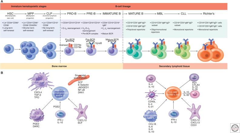
Patients with chronic lymphocytic leukemia can be divided into three categories: those who are minimally affected by the problem, often never requiring therapy; those that initially follow an indolent course but subsequently progress and require therapy; and those that from the point of diagnosis exhibit an aggressive disease necessitating treatment. Likewise, such patients pass through three phases: development of the disease, diagnosis, and need for therapy. Finally, the leukemic clones of all patients appear to require continuous input from the exterior, most often through membrane receptors, to allow them to survive and grow. This review is presented according to the temporal course that the disease follows, focusing on those external influences from the tissue microenvironment (TME) that support the time lines as well as those internal influences that are inherited or develop as genetic and epigenetic changes occurring over the time line. Regarding the former, special emphasis is placed on the input provided via the B-cell receptor for antigen and the C-X-C-motif chemokine receptor-4 and the therapeutic agents that block these inputs. Regarding the latter, prominence is laid upon inherited susceptibility genes and the genetic and epigenetic abnormalities that lead to the developmental and progression of the disease.


Figures

Figure 1.
Figure 1.
(Continued.) Microenvironment-supported B-lymphocyte development in patients with chronic lymphocytic leukemia (CLL). (A) Differentiation scheme assumed for CLL and Richter's transformation (RT) cells. Normal B lymphocytes progress through an ordered differentiation program that begins with hematopoietic stem cells (HSCs), proceeds through multipotent progenitor cells (MPPs), and then common lymphoid precursors (CLPs). After this point, commitment is made to the B-lymphocyte lineage. Pro-, pre-, and immature B cells differ based on the progressive rearrangements of IGHV, IGHD, and IGHJ genes. IGHD-IGHJ rearrangement is completed in pro-B cells, followed by IGHV-IGHD-IGHJ rearrangement in pre-B cells, and both IGHV-IGHD-IGHJ + IGLV-IGLJ rearrangements in immature B cells. Thus, antibodies that can create a complete B-cell receptor (BCR) exist from the pre-B cell–immature B cell interphase and at later stages of B-cell development (transitional, mature, etc.). Mutations in CLL HSCs and MPPs have been documented (Damm et al. 2014; Marsilio et al. 2018), and xenografting CLL HSCs/MPPs into severely immune-compromised mice (Kikushige et al. 2011) leads to a condition resembling monoclonal B-lymphocytosis (Rawstron et al. 2002a). If leukemia stem cells exist in CLL, HSCs, MPPs, CLPs, and pro-B cells carrying genetic abnormalities would be considered “pre-leukemic stem cells” and not “leukemic stem cells,” because they could not give rise to a CLL cell with the same IGHV-IGHD-IGHJ + IGLV-IGLJ rearrangements. The latter is relevant because, upon disease relapse, the emerging cells express a complete B-cell receptor (IGHV-IGHD-IGHJ + IGLV-IGLJ rearrangements) that indicates it is a member of the original CLL clone. Hence, we suggest the true leukemic stem cell in CLL resides at a differentiation stage at or beyond the pre-B cell–immature B cell interphase. CLL cells proliferate in secondary lymphoid organs and can transformation to Richter's cells, often as CLL-derived diffused large B-cell lymphoma (DLBCL) that have larger cells with prominent nuclei. (B) Microenvironmental signals associated with clonal expansion. (Left) Bone marrow microenvironment that supports normal and abnormal hematopoietic development. Bone marrow niche supports the differentiation and maturation of B cells. Immature HSCs and B cells are maintained and regulated by niche factors (i.e., CXCL12 and SCF produced by MSC), endothelial cell–derived signals (i.e., SCF, E-selectin, JAGGED1), and the progeny of HSCs including macrophages and megakaryocyte and regulatory T cells. (Right) The secondary lymphoid microenvironment sustains CLL and RT cells. CLL and RT cells receive growth promotion (e.g., IL-4, IL-21, CD40L, CCL19) and suppression (e.g., IL-10, IL-35, and PDL1-PD1 interaction) signals, as well as cell homing and retention cues (CXCL12, CXCL13, and CD31). Th2, T helper 2 cells; FDC, follicular dendritic cell; NK, natural-killer cell; MSC, mesenchymal stromal cell; Treg, T regulatory cell.
Figure 2.
Figure 2.
B-cell receptor (BCR) signaling pathway and the effects of kinase inhibition. Upon antigen interaction with the membrane IG component of the BCR, Lyn is activated and in turn phosphorylates CD79 on tyrosine residues—in particular, those in ITAM motifs. This permits binding of SYK and various adaptor molecules that initiate signaling that proceeds through key kinases including BTK, PI3Kδ, and AKT. (For a comprehensive review, see Slupsky [2014)].) The sites of action for kinase inhibitors are indicated: BTK (ibrutinib), PI3Kδ (idelalisib), PI3Kδ+γ (duvelisib), and SYK (fostamatinib).
Figure 3.
Figure 3.
CXCR4 signaling pathway and the effects of kinase inhibition. CXCR4 (C-X-C-motif chemokine receptor-4) is a G protein–coupled receptor (GPCR) that binds CXCL12/stromal-derived-factor-1 (SDF-1). CXCR4 signaling in response to CXCL12/SDF-1 mediates migration of circulating CLL cells. As a GPCR, CXCR4 engagement activates G protein–mediated signaling resulting in intracellular Ca2+ flux and subsequent activation of downstream pathways such as Ras and PI3Kδ. Activated JNK and PI3Kδ signaling lead to cell survival and migration. CXCR4 induces downstream signaling by several pathways such as those involving BTK and PLCγ2 signaling. Blocking CXCR4 signaling can be achieved by AMD3100 and plerixafor, which prevent CXCR4/SDF-1 binding, and by BTK inhibitor by ibrutinib. Other small molecular inhibitors like idelalisib and duvelisib target PI3K to block CXCR4 downstream signaling.
Figure 4.
Figure 4.
Life cycle of a chronic lymphocytic leukemia (CLL) B lymphocyte. In primary and secondary lymphoid tissues (bottom left) CLL B lymphocytes nestle, in a resting state, on survival-nurturing cells (e.g., nurse-like cells [Burger et al. 2000] or less well-defined, nonhematopoietic “stromal cells”). Docking is facilitated by several receptor–ligand interactions, including CXCL12-CXCR4. When cell division initiates, spontaneously or after stimulation via receptors (e.g., B-cell receptor [BCR] or Toll-like receptors [TLRs]), cells internalize CXCR4, detach, and migrate to structures resembling germinal centers (“proliferation centers” [Swerdlow et al. 1984; Schmid and Isaacson 1994; Bonato et al. 1998]), sites of CLL B-cell expansion. Activated/dividing cells up-regulate a number of surface proteins that promote interactions with T lymphocytes. These interactions up-regulate the DNA-mutating enzyme AID, which can cause mutations in genes genome-wide, leading to clonal evolution and possibly more aggressive disease. After expansion, some recently divided CLL cells exit the lymphoid tissue, entering the circulation bearing the CXCR4DimCD5Bright phenotype (“proliferative fraction” [Calissano et al. 2011]). Over time, the circulating cells express more CXCR4 and less CD5, eventually morphing into a CXCR4BrightCD5Dim resting fraction (RF) phenotype. These cells are best suited to follow a CXCL12/SDF1 gradient because of high levels of CXCR4, returning back to nutrient-rich niches in tissues and being rescued from apoptosis by IL-4 and likely other cytokines. Cells that cannot reenter tissues or do not arrive soon enough die. Once rescued, CLL cells proceed to a proliferation center to re-initiate the proliferative process and potential further clonal evolution or dock on a stromal element, where they again reside in the resting state.

References

    1. Acharya UH, Dhawale T, Yun S, Jacobson CA, Chavez JC, Ramos JD, Appelbaum J, Maloney DG. 2019. Management of cytokine release syndrome and neurotoxicity in chimeric antigen receptor (CAR) T cell therapy. Expert Rev Hematol 12: 195–205. 10.1080/17474086.2019.1585238 - DOI - PubMed
    1. Adams JM, Cory S. 2018. The BCL-2 arbiters of apoptosis and their growing role as cancer targets. Cell Death Differ 25: 27–36. 10.1038/cdd.2017.161 - DOI - PMC - PubMed
    1. Agathangelidis A, Darzentas N, Hadzidimitriou A, Brochet X, Murray F, Yan XJ, Davis Z, van Gastel-Mol EJ, Tresoldi C, Chu CC, et al. 2012. Stereotyped B-cell receptors in one-third of chronic lymphocytic leukemia: A molecular classification with implications for targeted therapies. Blood 119: 4467–4475. 10.1182/blood-2011-11-393694 - DOI - PMC - PubMed
    1. Agathangelidis A, Ljungström V, Scarfò L, Fazi C, Gounari M, Pandzic T, Sutton L-A, Stamatopoulos K, Tonon G, Rosenquist R, et al. 2018. Highly similar genomic landscapes in monoclonal B-cell lymphocytosis and ultra-stable chronic lymphocytic leukemia with low frequency of driver mutations. Haematologica 103: 865–873. 10.3324/haematol.2017.177212 - DOI - PMC - PubMed
    1. Agathangelidis A, Psomopoulos F, Stamatopoulos K. 2019. Stereotyped B cell receptor immunoglobulins in B cell lymphomas. Methods Mol Biol 1956: 139–155. 10.1007/978-1-4939-9151-8_7 - DOI - PubMed

MeSH terms

Substances

LinkOut - more resources