Review
. 2020 Mar 28;21(7):2346.
doi: 10.3390/ijms21072346.

Understanding MAPK Signaling Pathways in Apoptosis

Affiliations
Review

Understanding MAPK Signaling Pathways in Apoptosis

Jicheng Yue et al. Int J Mol Sci. .

Abstract

MAPK (mitogen-activated protein kinase) signaling pathways regulate a variety of biological processes through multiple cellular mechanisms. In most of these processes, such as apoptosis, MAPKs have a dual role since they can act as activators or inhibitors, depending on the cell type and the stimulus. In this review, we present the main pro- and anti-apoptotic mechanisms regulated by MAPKs, as well as the crosstalk observed between some MAPKs. We also describe the basic signaling properties of MAPKs (ultrasensitivity, hysteresis, digital response), and the presence of different positive feedback loops in apoptosis. We provide a simple guide to predict MAPKs' behavior, based on the intensity and duration of the stimulus. Finally, we consider the role of MAPKs in osmostress-induced apoptosis by using Xenopus oocytes as a cell model. As we will see, apoptosis is plagued with multiple positive feedback loops. We hope this review will help to understand how MAPK signaling pathways engage irreversible cellular decisions.

Keywords: ERK; JNK; MAPK; apoptosis; caspases; feedback loops; oocytes; p38; protein kinases; signaling.


Conflict of interest statement

The authors declare no conflict of interest.

Figures

Figure 1
Figure 1
JNK (C-Jun N-terminal kinase) substrates that regulate the mitochondrial apoptotic pathway. JNK can have pro-apoptotic effects through direct phosphorylation of Bcl-2 (B-cell lymphoma 2) family members. The phosphorylation of Bax (Bcl-2-associated X protein) will activate the pro-apoptotic activity of this protein, whereas the phosphorylation of Bcl-2 and Mcl-1 (myeloid cell leukemia 1) will suppress their anti-apoptotic function. Indeed, the direct phosphorylation and activation of Bad (Bcl-2-antagonist of cell death), Bim (Bcl-2-like protein 11), or Bmf (Bcl-2-modifying factor) will inhibit the antiapoptotic effect of Bcl-2. In addition, JNK phosphorylation of 14-3-3 protein induces the release of pro-apoptotic proteins Bax and Bad. JNK phosphorylation of the E3 ubiquitin ligase iTCH (itchy homolog) promotes the degradation of caspase-8 inhibitor c-FLIP (cellular FLICE (FADD-like IL-1β converting enzyme) inhibitory protein), therefore promoting caspase-8 activation and Bid (BH3 interacting-domain death agonist) proteolysis (tBid). JNK, activated by TNFα (tumor necrosis factor alfa), also promotes apoptosis through proteolysis of Bid (jBid) by an unknown protease (X). All these modifications in the Bcl-2 family members induce the release of cytochrome c and/or Smac/DIABLO (second mitochondria-derived activator of caspases/direct inhibitor of apoptosis protein-binding protein with low pI) from the mitochondria and subsequent caspase activation. Symbols used: activation (), inhibition (―•). Pro-apoptotic members of the Bcl-2 family are represented in red and anti-apoptotic members in blue.
Figure 2
Figure 2
Basic properties of MAPK (mitogen-activated protein kinase) signaling pathways. (A) Ultrasensitivity: A small increase in the stimulus produces a very large response in MAPK activity. Graphically, this is illustrated with a sigmoid curve, implying a threshold level for MAPK activation. Several biochemical mechanisms can explain this behavior. (B) Hysteresis: MAPKs in front of a certain stimulus, and under certain conditions, present sustained activation even when the stimulus has disappeared but under different conditions or stimulus, could return to basal levels (no hysteresis). (C) Digital response: single cells exposed to a certain dose of stimulus will present an initial homogeneous activation of MAPK (20% activity, graded response), which could be converted into an all-or-none response (0% or 100% activity, digital response) at a later time. The generation of digital responses in stress sensors depends on time, threshold levels of activation, ultrasensitivity, and the presence of positive feedback loops. (D) Positive feedback-loop: An initial stimulus will activate X, which in turn will activate Y, which will feed back to the input X. These loops, that once engaged would be independent of the initial stimulus, are commonplace in irreversible biological processes.
Figure 3
Figure 3
Positive feedback loops in apoptosis. (A) Caspases, kinases, and Bcl-2 family members are functionally linked in positive feedback loops to induce cell death. (1) Cytochrome c release from the mitochondria, a meeting point in the cell death pathways, induces caspase activation. Caspase-9 and subsequent caspase-3 activation produces Bcl-2 proteolysis, which in turn promotes more cytochrome c release [108]. (2) Caspase-3 induces proteolysis and constitutive activation of MEKK1 (MAPK/ERK kinase kinase 1), that promotes cytochrome c release and caspase-9/-3 activation though JNK/p38 activation [119,120,121]. (3 and 4) Caspase-3 [110,111] or caspase-8 [109] induce Bid proteolysis (tBid), which in turn promotes cytochrome c release and caspase activation. (5) Caspase-3 is activated by caspase-9, which in turn can induce proteolysis and full activation of caspase-9 [112,113]. (B) ROS (reactive oxygen species) generation and ASK1 (apoptosis signal-regulating kinase 1) activation are sustained by multiple positive feedback loops. (1) NADPH oxidase generates ROS and activates ASK1 and JNK, which in turn activates NADPH oxidase [122,123]. (2 and 3) JNK and/or p38 activation, induced by ROS, produces p53 activation, which in turn induces ROS generation [27,124]. (4) Sustained activation of p38 increases ROS production, which in turn activates ASK1 and p38 [125,126]. (5) ASK1 activation induces the accumulation of Daxx protein, which in turn further activates ASK1 [129]. (6) ASK1 induces transcription of cyclin D1 through AP-1 (activator protein 1) activation, which in turn increases ASK1 levels via the Rb-E2F (retinoblastoma-E2F transcription factor) pathway [130]. (7) Ceramide activates the ASK1 and JNK signaling pathway, which in turn activates neutral sphingomyelinase (nSMase), thus increasing ceramide production [131,132]. The loops represented in the figure do not necessarily take place in the same cell and at the same time. Some loops are activated by a specific stimulus or in a certain cell type. Symbols used: activation (), inhibition (―•).
Figure 4
Figure 4
A model for osmostress-induced apoptosis. In Xenopus oocytes, hyperosmotic shock induces rapid calpain activation, Smac/DIABLO release from the mitochondria, cleavage of small amounts of Bid by an unknown protease, and MAPK activation (JNK1-1, JNK1-2, and p38). Low amounts of cytochrome c are released from the mitochondria at this early stage (0–1 h), but they are not sufficient to activate caspase-3. Stress protein kinases (JNKs and p38) would act as cellular sensors to evaluate the stressful situation. MAPKs can engage, by early phosphorylation of some substrates (A, B, C), a protective response. Mitochondria integrates the information received by stress sensors at this early stage, where the cell could recover if the stress does not persist or gets weaker. However, sustained and increased activation of MAPKs will lead to late phosphorylation and activation of pro-apoptotic substrates (X, Y, Z), including Bcl-2 family members. A marked release of cytochrome c will promote caspase-3 activation (2 h), which in turn would induce more calpain activation, cleavage of JNK1-2 and Bid, and p38 activation. These events promote additional cytochrome c release and caspase-3 activation in multiple positive feedback loops, resulting in an irreversible apoptotic process (2–4 h). The blue color in the arrows represents early responses to stress in the reversible phase of apoptosis, and the red color represents late responses in the irreversible phase.
Figure 5
Figure 5
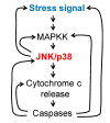
MAPKs in apoptosis: A simplified model. An input, represented by a stress signal (Ca2+, ROS, osmotic shock, death receptor, etc.), will activate a MAPK cascade, which in turn feedbacks on the initial input/cascade to achieve higher MAPK activation. After a threshold of MAPK activation is surpassed, additional downstream targets are activated, promoting cytochrome c release and caspase activation. Other positive feedback loops, engaged downstream of MAPKs, will keep sustained and high levels of activation of JNK/p38 and caspases, assuring the irreversibility of the apoptotic process.

References

    1. Ray L.B., Sturgill T.W. Rapid stimulation by insulin of a serine/threonine kinase in 3T3-L1 adipocytes that phosphorylates microtubule-associated protein 2 in vitro. Proc. Natl. Acad. Sci. USA. 1987;84:1502–1506. doi: 10.1073/pnas.84.6.1502. - DOI - PMC - PubMed
    1. Ray L.B., Sturgill T.W. Insulin-stimulated microtubule-associated protein kinase is phosphorylated on tyrosine and threonine in vivo. Proc. Natl. Acad. Sci. USA. 1988;85:3753–3757. doi: 10.1073/pnas.85.11.3753. - DOI - PMC - PubMed
    1. Boulton T.G., Yancopoulos G.D., Gregory J.S., Slaughter C., Moomaw C., Hsu J., Cobb M.H. An insulin-stimulated protein kinase similar to yeast kinases involved in cell cycle control. Science. 1990;249:64–67. doi: 10.1126/science.2164259. - DOI - PubMed
    1. Boulton T.G., Nye S.H., Robbins D.J., Ip N.Y., Radziejewska E., Morgenbesser S.D., DePinho R.A., Panayotatos N., Cobb M.H., Yancopoulos G.D. ERKs: A family of protein-serine/threonine kinases that are activated and tyrosine phosphorylated in response to insulin and NGF. Cell. 1991;65:663–675. doi: 10.1016/0092-8674(91)90098-J. - DOI - PubMed
    1. Davis R.J. Signal transduction by the JNK group of MAP kinases. Cell. 2000;103:239–252. doi: 10.1016/S0092-8674(00)00116-1. - DOI - PubMed

Substances