. 2020 Mar 30;11(1):1606.
doi: 10.1038/s41467-020-15459-7.

Structural insights into tetraspanin CD9 function

Affiliations

Structural insights into tetraspanin CD9 function

Rie Umeda et al. Nat Commun. .

Abstract

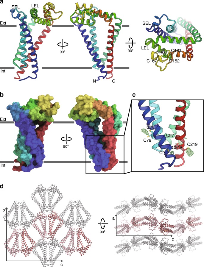
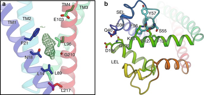
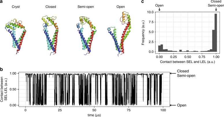
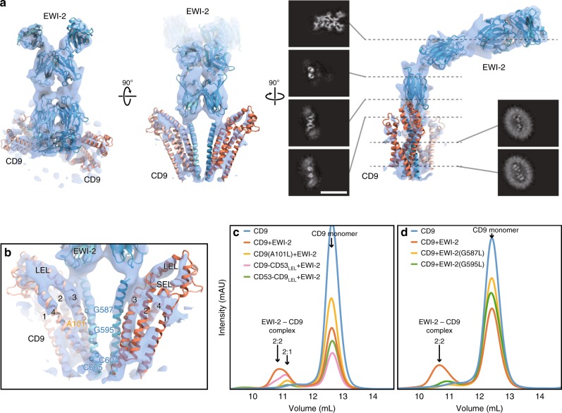
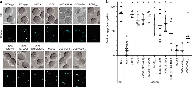
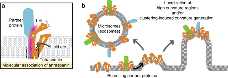
Tetraspanins play critical roles in various physiological processes, ranging from cell adhesion to virus infection. The members of the tetraspanin family have four membrane-spanning domains and short and large extracellular loops, and associate with a broad range of other functional proteins to exert cellular functions. Here we report the crystal structure of CD9 and the cryo-electron microscopic structure of CD9 in complex with its single membrane-spanning partner protein, EWI-2. The reversed cone-like molecular shape of CD9 generates membrane curvature in the crystalline lipid layers, which explains the CD9 localization in regions with high membrane curvature and its implications in membrane remodeling. The molecular interaction between CD9 and EWI-2 is mainly mediated through the small residues in the transmembrane region and protein/lipid interactions, whereas the fertilization assay revealed the critical involvement of the LEL region in the sperm-egg fusion, indicating the different dependency of each binding domain for other partner proteins.


Conflict of interest statement

The authors declare no competing interests.

Figures

Fig. 1
Fig. 1. Crystal structure of human CD9.
a Overall structure of human CD9, viewed from the membrane plane (left and middle) and from the extracellular side (right). The transmembrane helices and the two extracellular loops (SEL and LEL) are labeled. Cys152-Cys181 and Cys153-Cys167 form disulfide bonds. b Surface representation of CD9, colored according to a. c Palmitoylation of the cytoplasmic cysteine residues. Green meshes show FoFc densities contoured at 2.5 σ, indicating the palmitoylation of the cysteine residues on the cytoplasmic end of the four transmembrane helices. d Crystal packing of CD9, showing the curvature generation by the asymmetric shape of the CD9 molecules. Pairs of CD9 protomers are aligned up and down alternatively along the c-axis, resulting in the wavy lipid layers of the LCP crystal.
Fig. 2
Fig. 2. Central hydrophilic pocket and extracellular loops.
a The central cavity is rendered hydrophilic by the conserved residues, Asn18 and Glu103. The critical residue for cholesterol binding, Glu219 in CD81, is replaced by Gly210 in CD9 (Supplementary Fig. 1). The FoFc map contoured at 2.5 sigma shows an unknown density within the cavity, probably derived from the monoolein present during the lipidic cubic phase crystallization. b The central cavity is sealed on the extracellular side by the association between the SEL and LEL. The residues involved in the interactions are shown, and the hydrogen bonding interactions are indicated by yellow dotted lines.
Fig. 3
Fig. 3. MD simulation of CD9.
a Structural comparison between the crystal structure (Cryst) and the three major conformations in the MD simulation (Closed, Semi-open, and Open). b Time course of the contacts between the SEL and LEL in the reconstructed 100 μs trajectory. The value of 1 corresponds to the shorter distance between the SEL and LEL, and the value of 0 corresponds to the longer distance between the SEL and LEL. For the detailed definition of the contacts between the SEL and LEL, see Methods section. c Distribution of the contacts between the SEL and LEL in the longer reconstructed trajectory, corresponding to the 15 ms time scale.
Fig. 4
Fig. 4. Cryo-EM structure of CD9 in complex with EWI-2.
a Cryo-EM density map of the Fab-CD9-EWI-2 complex. The density for the Fab fragment was somewhat disordered (Supplementary Fig. 6). Homology models of the Ig-like domains of EWI-2 were constructed with the PHYRE2 server and roughly fitted into the density. The TM helices and the two extracellular loops (SEL and LEL) of the CD9 crystal structure are separately fitted into the density. The density for the detergent micelle is omitted for clarity. Black panels show sections of the density, demonstrating the C2 symmetry in the TM region and the proximal Ig-like domain. Scale bar, 100 Å. b Close-up view of the TM region. CD9 adopts a semi-open conformation with a slight rearrangement of the LEL, as compared to the crystal structure (Supplementary Fig. 6). The residues in the EWI-2 membrane-spanning region were modeled according to the mutant analyses in Fig. 2c, d. c, d Complex formation assays for CD9 mutants (c) and EWI-2 mutants (d). GFP-labeled CD9 and EWI-2 were co-expressed in HEK cells, and complex formation was analyzed by FSEC. The slight peak shift toward the smaller molecular weight indicates the complex formation with 2:1 stoichiometry.
Fig. 5
Fig. 5. Sperm–egg fusion complementation assay.
a Functional complementation of Cd9 KO mouse eggs by mRNA injection. The mRNA, encoding the WT or mutants of mouse CD9, human CD9, or chimeric constructs between human CD9 and CD53, was injected into the mouse Cd9 KO eggs. The fusion of mouse spermatozoa was visualized by the transfer of DNA dye from the eggs to the sperm nuclei. Asterisks indicate unfertilized eggs. Scale bar, 50 µm. b Fertilization rates in the sperm–egg fusion assay. The averaged fertilization rates of repetitive experiments are shown with error bar of standard deviation. Each dot represents one experiment. Asterisks indicate significant differences from the Cd9 KO eggs (two-tailed, unpaired Student’s t-test, *P < 0.005). The number of experiments and eggs are indicated in Supplementary Fig. 8c and also provided as a Source Data file.
Fig. 6
Fig. 6. Hypothetical model of the tetraspanin function.
a The molecular association of tetraspanin and its partner proteins is mediated through both the TM and LEL regions, but its dependency is different among the partner proteins. Lipid binding to the central cavity could modulate the molecular association by affecting the LEL conformation. b The highly asymmetric shape of the tetraspanins suggests their localization at high curvature regions and/or clustering-induced curvature generation. Partner proteins should also be recruited and sorted to the respective regions, such as the microvilli tips and microsomes (exosomes).

References

    1. Charrin S, Jouannet S, Boucheix C, Rubinstein E. Tetraspanins at a glance. J. Cell Sci. 2014;127:3641–3648. doi: 10.1242/jcs.154906. - DOI - PubMed
    1. Reimann R, Kost B, Dettmer J. Tetraspanins in plants. Front. Plant Sci. 2017;8:545. doi: 10.3389/fpls.2017.00545. - DOI - PMC - PubMed
    1. Huang S, et al. The phylogenetic analysis of tetraspanins projects the evolution of cell–cell interactions from unicellular to multicellular organisms. Genomics. 2005;86:674–684. doi: 10.1016/j.ygeno.2005.08.004. - DOI - PubMed
    1. Hemler ME. Tetraspanin proteins mediate cellular penetration, invasion, and fusion events and define a novel type of membrane microdomain. Annu. Rev. Cell Dev. Biol. 2003;19:397–422. doi: 10.1146/annurev.cellbio.19.111301.153609. - DOI - PubMed
    1. Stuck MW, Conley SM, Naash MI. PRPH2/RDS and ROM-1: historical context, current views and future considerations. Prog. Retinal Eye Res. 2016;52:47–63. doi: 10.1016/j.preteyeres.2015.12.002. - DOI - PMC - PubMed

Publication types

LinkOut - more resources