. 2020 Mar 13:11:268.
doi: 10.3389/fphar.2020.00268. eCollection 2020.

Rosiglitazone Protects Endothelial Cells From Irradiation-Induced Mitochondrial Dysfunction

Affiliations

Rosiglitazone Protects Endothelial Cells From Irradiation-Induced Mitochondrial Dysfunction

Bjorn Baselet et al. Front Pharmacol. .

Abstract

Background and purpose: Up to 50-60% of all cancer patients receive radiotherapy as part of their treatment strategy. However, the mechanisms accounting for increased vascular risks after irradiation are not completely understood. Mitochondrial dysfunction has been identified as a potential cause of radiation-induced atherosclerosis.

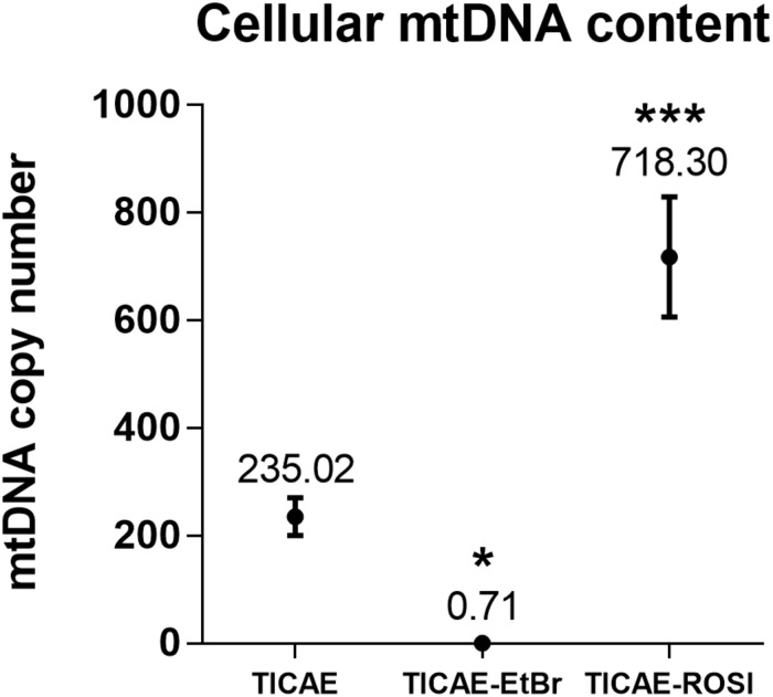
Materials and methods: Assays for apoptosis, cellular metabolism, mitochondrial DNA content, functionality and morphology were used to compare the response of endothelial cells to a single 2 Gy dose of X-rays under basal conditions or after pharmacological treatments that either reduced (EtBr) or increased (rosiglitazone) mitochondrial content.

Results: Exposure to ionizing radiation caused a persistent reduction in mitochondrial content of endothelial cells. Pharmacological reduction of mitochondrial DNA content rendered endothelial cells more vulnerable to radiation-induced apoptosis, whereas rosiglitazone treatment increased oxidative metabolism and redox state and decreased the levels of apoptosis after irradiation.

Conclusion: Pre-existing mitochondrial damage sensitizes endothelial cells to ionizing radiation-induced mitochondrial dysfunction. Rosiglitazone protects endothelial cells from the detrimental effects of radiation exposure on mitochondrial metabolism and oxidative stress. Thus, our findings indicate that rosiglitazone may have potential value as prophylactic for radiation-induced atherosclerosis.

Keywords: cardiovascular disease; endothelial cells; ionizing radiation; mitochondria; rosiglitazone.


Figures

FIGURE 1
FIGURE 1
Irradiation decreases TICAE cell mitochondrial abundance. (A) The graph shows mitochondrial GFP content per cell over time in sham-irradiated and 2 Gy-irradiated TICAE cells, determined using live cell imaging and normalized according to masked cell count based on bright field images. P-values are indicated as colored blocks (N = 3, n = 12). (B) Representative images of 0 Gy 48 h after irradiation (left) and a 2 Gy 48 h after irradiation (right). Bar = 300 μm. Statistics were performed by a multiple comparison two-way ANOVA with Bonferroni post hoc test.
FIGURE 2
FIGURE 2
Validation of TICAE-EtBr and TICAE-ROSI models. mtDNA copy number in untreated TICAE cells (control) and in TICAE-EtBr and TICAE-ROSI cells (N = 3, n = 12). *P < 0.05 and ***P < 0.001 by one-way ANOVA with Tukey post hoc test.
FIGURE 3
FIGURE 3
Rosiglitazone treatment protects TICAE cells against irradiation. Graphs show the apoptotic index of TICAE, TICAE-EtBr and TICAE-ROSI cells 98 h after a single 2 Gy irradiation, using annexin V (left) and activated caspase 3/7 staining (right) (N = 2, n = 7–14). *P < 0.05, **P < 0.01, ****P < 0.0001 by one-way ANOVA with Tukey post hoc test.
FIGURE 4
FIGURE 4
Irradiation decreases TICAE cell respiration but increases their respiration capacity. The rates of basal respiration, respiration linked to ATP production, maximal respiration and spare respiratory capacity were measured using Seahorse oximetry in TICAE, TICAE-EtBr and TICAE-ROSI cells 24 h after sham (●) or 2 Gy X-ray (formula image) exposure (N = 3, n = 32–64). *P < 0.05, ****P < 0.0001 by two-way ANOVA with Bonferroni post hoc test.
FIGURE 5
FIGURE 5
Irradiation minimally affects ATP levels in endothelial cells. Measurements of ATP concentrations in TICAE, TICAE-EtBr and TICAE-ROSI cells at increasing times after a sham or a 2 Gy X-ray irradiation (N = 2, n = 24). ****P < 0.0001 using two-way ANOVA with Bonferroni post hoc test.
FIGURE 6
FIGURE 6
Endothelial cells with dysfunctional mitochondria have difficulties to cope with redox stress upon irradiation. (A) Intracellular ROS measurements in TICAE, TICAE-EtBr and TICAE-ROSI cells 24 h after a sham or a 2 Gy X-ray irradiation (N = 3, n = 32). (B) Same as A but basal ROS were measured after exposing the cells to 20 μM tert-butyl peroxide (tBHP) (N = 3, n = 32). (C) Mitochondrial membrane potential of TICAE, TICAE-EtBr and TICAE-ROSI cells 24 h after a sham or a 2 Gy X-ray irradiation (N = 3, n = 32). ***P < 0.005, ****P < 0.0001 by two-way ANOVA with Bonferroni post hoc test.
FIGURE 7
FIGURE 7
Irradiation alters mitochondrial morphology in endothelial cells. (A) Mitochondrial surface relative to cell surface was measured in TMRM-stained TICAE, TICAE-EtBr and TICAE-ROSI cells 24 h after a sham or a 2 Gy X-ray irradiation (N = 3, n = 32). (B) As in A, but measuring mitochondrial circularity (N = 3, n = 32). (C) Representative TEM pictures of TICAE, TICAE-EtBr and TICAE-ROSI cells 24 h after a sham or a 2 Gy X-ray irradiation. Black arrows point at normal mitochondria and white arrows at abnormal mitochondria. Bars = 5 μm. *P < 0.05, ****P < 0.0001 by two-way ANOVA with Bonferroni post hoc test.

References

    1. Ahmad T., Aggarwal K., Pattnaik B., Mukherjee S., Sethi T., Tiwari B. K., et al. (2013). Computational classification of mitochondrial shapes reflects stress and redox state. Cell Death Dis. 4:e461. 10.1038/cddis.2012.213 - DOI - PMC - PubMed
    1. Bakkal B. H., Gultekin F. A., Guven B., Turkcu U. O., Bektas S., Can M. (2013). Effect of ozone oxidative preconditioning in preventing early radiation-induced lung injury in rats. Braz. J. Med. Biol. Res. 46 789–796. 10.1590/1414-431x20132856 - DOI - PMC - PubMed
    1. Belikova N. A., Glumac A., Rafikov R., Jiang J., Greenberger J. S., Kagan V. E., et al. (2009). Radioprotection by short-term oxidative preconditioning: role of manganese superoxide dismutase. FEBS Lett. 583 3437–3442. 10.1016/j.febslet.2009.10.013 - DOI - PMC - PubMed
    1. Bhattacharya S., Asaithamby A. (2016). Ionizing radiation and heart risks. Semin. Cell Dev. Biol. 58 14–25. 10.1016/j.semcdb.2016.01.045 - DOI - PubMed
    1. Bogliolo M., Izzotti A., De Flora S., Carli C., Abbondandolo A., Degan P. (1999). Detection of the ‘4977 bp’ mitochondrial DNA deletion in human atherosclerotic lesions. Mutagenesis 14 77–82. 10.1093/mutage/14.1.77 - DOI - PubMed

LinkOut - more resources