. 2020;8(2-3):67-78.
doi: 10.1080/21678707.2020.1735352. Epub 2020 Mar 2.

Monitoring progression of retinitis pigmentosa: current recommendations and recent advances

Affiliations

Monitoring progression of retinitis pigmentosa: current recommendations and recent advances

Moreno Menghini et al. Expert Opin Orphan Drugs. 2020.

Abstract

Introduction: Retinitis pigmentosa (RP) is the most common form of inherited retinal degenerations with an estimated prevalence of 1 in 4,000 and more than 1 million individuals affected worldwide. With the introduction of the first retinal gene therapy in 2017 the importance of understanding the mechanisms of retinal degeneration and its natural progression has shifted from being of academic interest to being of pivotal for the development of new therapies.

Areas covered: This review covers standard and innovative diagnostic techniques and complementary examinations needed for the evaluation and treatment of RP. It includes chapters on the assessment of visual function, retinal morphology, and genotyping.

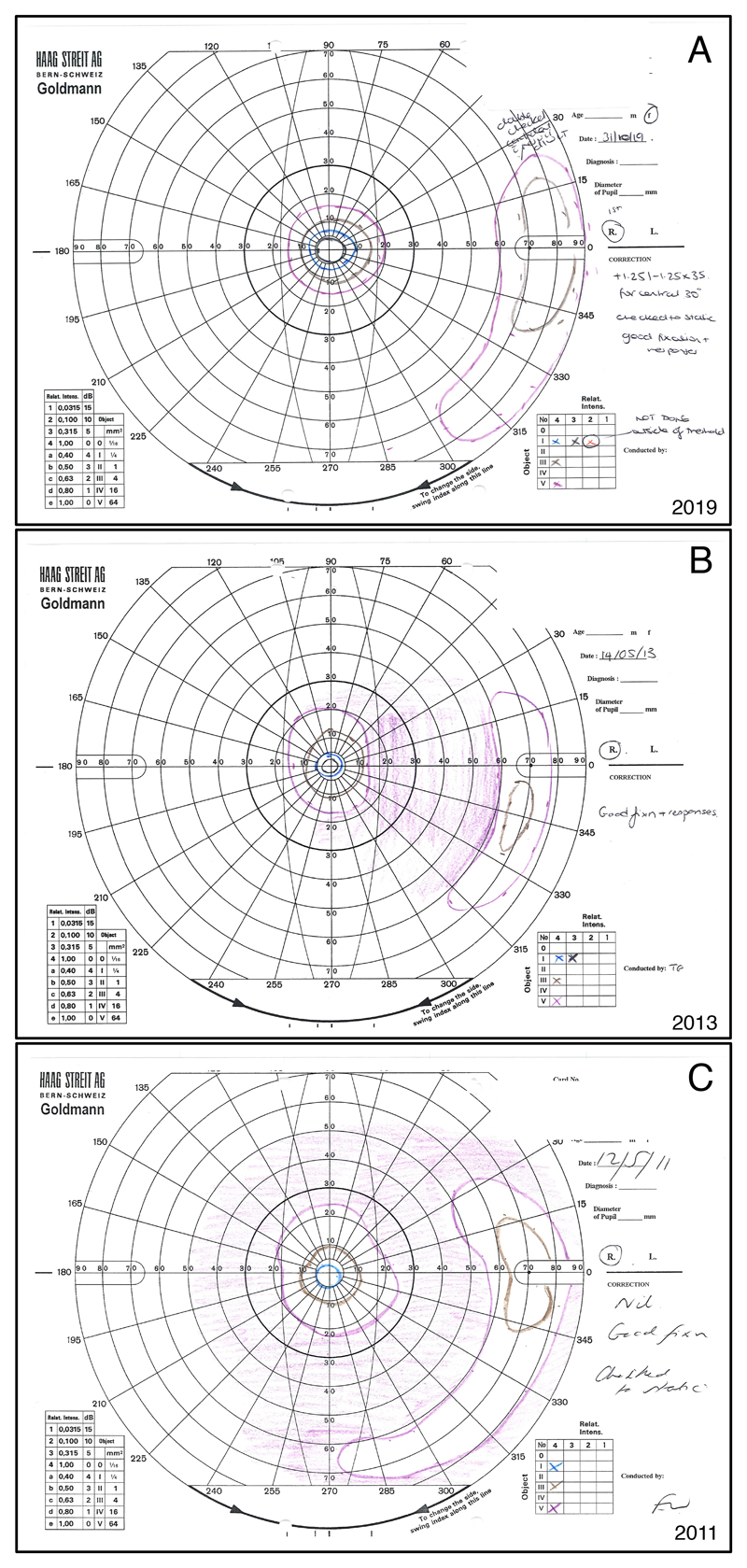
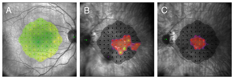
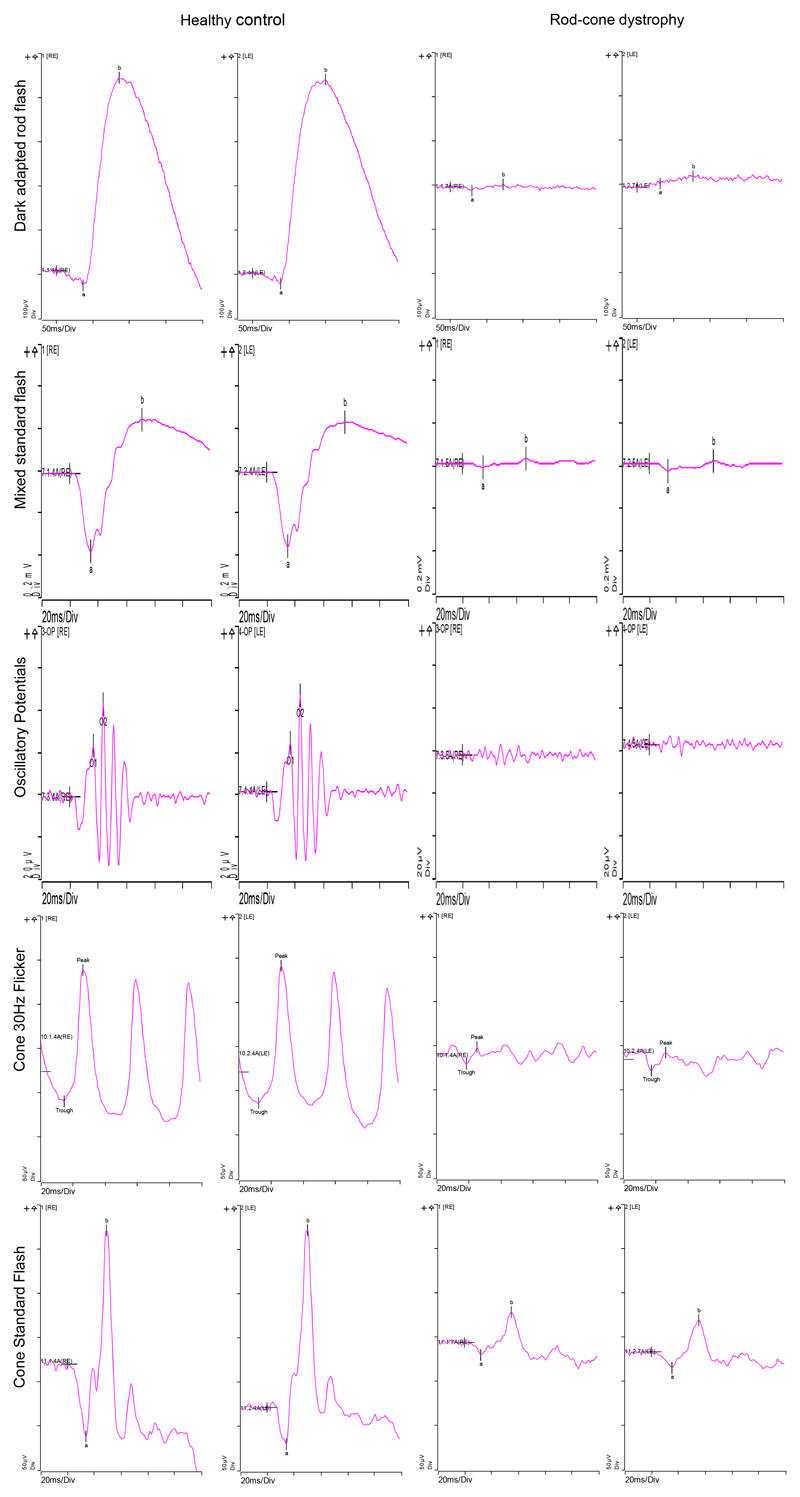
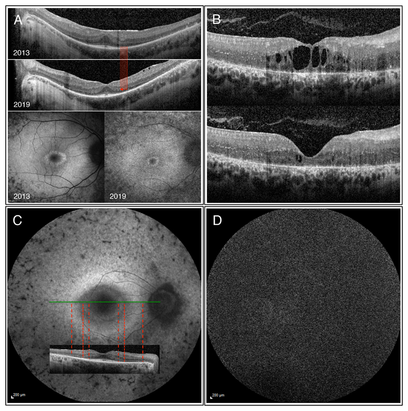
Expert opinion: Monitoring the progression of RP can best be achieved by combining assessments of both visual function and morphology. Visual acuity testing using ETDRS charts should be complemented by low-luminance visual acuity and colour vision tests. Assessment of the visual field can also be useful in less advanced cases. In those with central RP involvement measuring retinal sensitivity using microperimetry is recommended. Retinal morphology is best assessed by OCT and autofluorescence. Genetic testing is pivotal as it contributes to the pathophysiological understanding and can guide clinical management as well as identify individuals that could benefit from retinal gene therapy.

Keywords: Autofluorescence; ETDRS letters; IRD; OCT; RP; genotyping; inherited retinal degeneration; microperimetry; optical coherence tomography; retinitis pigmentosa.


Figures

Figure 1
Figure 1
Goldmann visual fields of the right eye of a 37 year-old female with PDE6B associated autosomal recessive RP. Figure 1A shows the most recent visual field from 2019, while 1B is from 2013, and 1C from 2011. The normal Goldmann visual field usually reaches about 90° temporally, 50° nasally, and 50-60° superiorly and inferiorly for the largest and brightest stimulus. All fields show a concentric constriction with a peripheral remnant of visual field, which is typical for RP. Significantly increased constriction and decrease in the size of the peripheral remnant can be noted from 2011 to 2013. However, the visual field from 2019 seems to not have worsened in comparison to the previous exams. Indeed, both the central island and the peripheral remnant appear larger. A test-retest variability of up to 20% even when using a single experienced operator has been known for Goldmann visual fields, and might explain the apparent improvement in visual field seen in this patient. The clinical improvement seen in the cystoid macular oedema between 2013 and 2019 (not shown) could however have contributed to a real improvement in visual field sensitivity in this case. The purple lines represent the Goldmann stimulus V4e. The Roman number indicates the stimulus size of 64mm2 for V. The Arabic number and the lower case letter indicate the light intensity of 1000 apostilb (315 cd/m2). Brown = III4e (4mm2, 1000 asb); blue = I4e (1mm2, 1000 asb); black = I3e (1mm2, 315 asb).
Figure 2
Figure 2
Standard 10-2 grid consisting of 68 retinal points arranged in a Cartesian pattern covering the central 20°. The threshold sensitivity value at each retinal location is colour coded and shown as an overlay on the near-infrared image. A) Macular sensitivity heat map of a healthy 27 year-old male. Green indicates normal sensitivity (maximum 36dB). B and C) Macular sensitivity heat map of a 38 year-old male with RPGR X-linked RP taken two years apart. In 2017 (B) the patient still showed few retinal points of nearly normal sensitivity, while two years later (C) both the central visual field constriction, and sensitivity threshold had worsened.
Figure 3
Figure 3
ISCEV Standard ERG of a 10 year-old boy with a strong family history of PRPF31 associated RP. Normal values of a healthy control are shown on the left columns. The right columns show the ERG of the 10 year-old boy. The dark-adapted rod flash (0.01 cd*s/m2) shows barely measurable components. The mixed standard flash (3 cd*s/m2) reveals reduced amplitudes, whilst peak times are normal. The third row shows barely recordable dark-adapted (3 cd*s/m2) oscillatory potentials. The light-adapted 30 Hz flicker ERG and the cone standard flash (3 cd*s/m2) both show markedly reduced responses. In summary, the ERG confirms rod and cone dysfunction in both eyes. Overall rods seem to be more affected than cones at this stage.
Figure 4
Figure 4
OCT and autofluorescence images of various patients with RP. A) Images of the right eye of a 28 year-old male with autosomal recessive RP (compound heterozygous mutations in DFNB31 and USH2A found). Significant decrease in the width of both the ELM and EZ paralleled by diminishing foveal hypoautofluorescence can be observed between 2013 and 2019. Visual acuity in 2019 was 6/7.5, which translates to 20/25 (20 ft) or 0.8 (decimal) Snellen equivalent. B) OCT images of the right eye of a 26 year-old female patient with CRB1 associated RP showing clinically meaningful improvement of the cystoid macular oedema upon systemic acetazolamide therapy with an increase in visual acuity from 3/60 (20/400, 0.05) to 6/38 (20/125, 0.16). C) Autofluorescence and OCT images of the right eye of a 61 year-old male with PRPF8 associated autosomal dominant RP. The inner hyperautofluorescent circle shows good correlation with the loss in ELM, while the outer larger hyperautofluorescent ring correlates with retinal area of absent ELM and highly thinned ONL. D) Short-wavelength autofluorescence image of a 48 y/o male patient with biallelic RPE65 (c.11+5G>A, c.1543C>T) showing the characteristic absent autofluorescence.

References

    1. Berger W, Kloeckener-Gruissem B, Neidhardt J. The molecular basis of human retinal and vitreoretinal diseases. [cited 2019 Sep 25];Prog Retin Eye Res. 2010 29:335–375. [Internet]. Available from: https://www.sciencedirect.com/science/article/pii/S135094621000025X. - PubMed
    1. Hartong DT, Berson EL, Dryja TP. Retinitis pigmentosa. [cited 2019 Sep 25];Lancet. 2006 368:1795–1809. [Internet]. Available from: https://linkinghub.elsevier.com/retrieve/pii/S0140673606697407. - PubMed
    1. Pagon RA. Retinitis pigmentosa. [cited 2019 Sep 25];Surv Ophthalmol. 1988 33:137–177. [Internet]. Available from: https://linkinghub.elsevier.com/retrieve/pii/0039625788900859. - PubMed
    1. Verbakel SK, Van Huet RAC, Boon CJF, et al. Progress in Retinal and Eye Research Non-syndromic retinitis pigmentosa. Prog Retin Eye Res. 2018;66:1–30. doi: 10.1016/j.preteyeres.2018.03.005. [Internet]. - DOI - PubMed
    1. Prokisch H, Hartig M, Hellinger R, et al. A Population-Based Epidemiological and Genetic Study of X-Linked Retinitis Pigmentosa. [cited 2019 Sep 25];Investig Opthalmology Vis Sci. 2007 48:4012. doi: 10.1167/iovs.07-0071. [Internet]. - DOI - PubMed

LinkOut - more resources