Review
. 2020 Mar 13:10:322.
doi: 10.3389/fonc.2020.00322. eCollection 2020.

Autophagy Regulation by the Translation Machinery and Its Implications in Cancer

Affiliations
Review

Autophagy Regulation by the Translation Machinery and Its Implications in Cancer

Pilar Sarah Acevo-Rodríguez et al. Front Oncol. .

Abstract

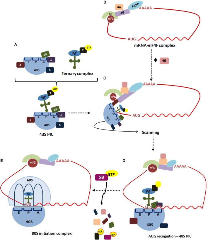
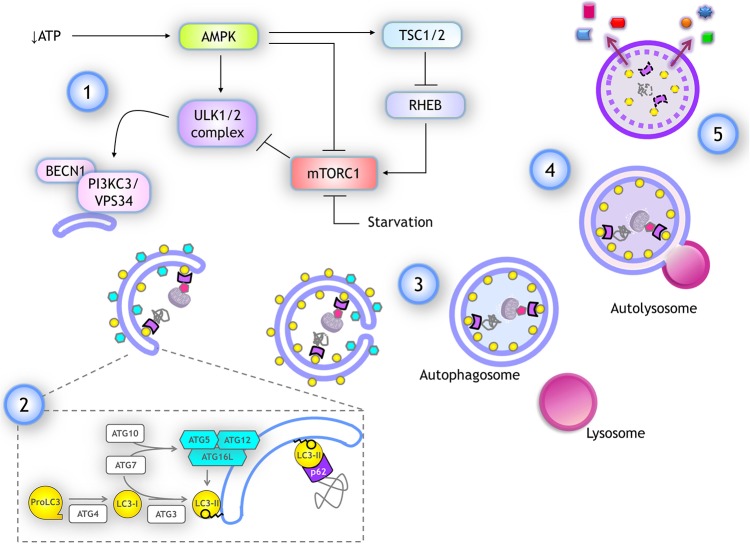
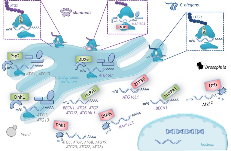
Various metabolic pathways and molecular processes in the cell act intertwined, and dysregulating the interplay between some of them may lead to cancer. It is only recently that defects in the translation process, i.e., the synthesis of proteins by the ribosome using a messenger (m)RNA as a template and translation factors, have begun to gain strong attention as a cause of autophagy dysregulation with effects in different maladies, including cancer. Autophagy is an evolutionarily conserved catabolic process that degrades cytoplasmic elements in lysosomes. It maintains cellular homeostasis and preserves cell viability under various stress conditions, which is crucial for all eukaryotic cells. In this review, we discuss recent advances shedding light on the crosstalk between the translation and the autophagy machineries and its impact on tumorigenesis. We also summarize how this interaction is being the target for novel therapies to treat cancer.

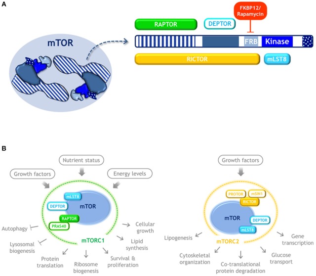
Keywords: ATG; PERK; autophagy; cancer; eIF2alpha; endoplasmic reticulum; mTOR; translation initiation.


Figures

Figure 1
Figure 1
Translation initiation in eukaryotes. Translation of most eukaryotic mRNAs is mediated by the eukaryotic initiation factors (eIFs). (A) This process begins when the free 40S ribosomal subunit, which is stabilized by eIF3 (3), eIF1 (1), eIF1A (1A), and eIF5 (5), binds to a ternary complex consisting of eIF2-GTP bound to an initiator Met-tRNAi, forming 43S pre-initiation complex (PIC). (B) Simultaneously, the cap structure (m7G) located at the 5′-end of an mRNA is recognized by the cap-binding protein, eIF4E (4E). The scaffold protein eIF4G (4G) performs simultaneous interactions with the cap-bound eIF4E, the ATP-dependent RNA-helicase eIF4A (4A) and PABP bound to poliA, circularizing the mRNA to form the mRNA-eIF4F complex. (C) The ribosome-bound eIF3 coordinates the recruitment of the 43S PIC to the mRNA 5'-UTR. The 43S PIC scans base-by-base the mRNA 5′-UTR to reach the AUG start codon, a process in which eIF4A, assisted by eIF4B (4B), unwinds secondary structures of the 5′-UTR. (D) Selection of the start codon establishes the open reading frame for mRNA decoding, and results in a 48S PIC with the Met-tRNAiMet and eIF1A tightly positioned within the P-site. (E) Then, a GTP-eIF5B complex promotes release of eIF1 and eIF5B, facilitating joining of a 60S ribosomal subunit to the 48S PIC to assemble an 80S initiation complex, which is ready to start the elongation step of translation.
Figure 2
Figure 2
mTOR kinase structure and complexes. (A) Schematic representation of mTOR kinase domains and its interacting proteins. mTOR possess 5 main domains (highlighted in blue). As an active form, mTOR dimerizes and may form two distinct complexes. mTORC1 is composed by three subunits that cooperate to phosphorylate substrates: mTOR, RAPTOR, and mLST8, and by the inhibitory subunits DEPTOR and PRAS40. Rapamycin forms a complex with FKBP12 that binds to mTOR and inhibits mTORC1 signaling. mTORC2 also contains mTOR, DEPTOR, and mLST8, but instead of RAPTOR it contains RICTOR, as well as the regulatory subunits mSIN1, and PROTOR. (B) mTORC1 and mTORC2 respond to distinct stimulus and control different cellular process. Color code: blue, mTOR kinase; cyan, components of both mTOR complexes; green, MTORC1 exclusive components; yellow, MTORC2 exclusive components.
Figure 3
Figure 3
Autophagy: overview and key molecular components. (1) Several stimuli promoting autophagy, like a drop in ATP, lead to AMPK activation, which stimulates autophagy by activating ULK1/2 complex and inhibiting mTORC1 through TSC1/2 activation, which in turn inactivates RHEB, a negative regulator of mTORC1. ULK1/2 complex activates the class III PI3K complex I by phosphorylating PIK3C3/VPS34. (2) For phagophore elongation, conjugation of ATG5-ATG12 complex is catalyzed by ATG7 and ATG10. ATG5-ATG12 covalently linked then interact with ATG16L forming a complex that is recruited at the phagophore. LC3 is proteolytically cleaved upon its translation by ATG4, producing LC3-I form. When autophagy is induced, LC3-I is covalently bound to phosphatidylethanolamine (PE) at the membrane of the phagophore. This reaction is catalyzed by ATG3 (E1-like) and ATG7 (E2-like) again, while ATG12-ATG5/ATG16L complex already recruited at the phagophore surface functions as an E3-like enzyme. Lipidated LC3-I is named LC3-II and it remains anchored to the elongating phagophore. LC3-II associates to both inner and outer membranes of the phagophore in expansion. Cargo is recognized by adaptor proteins like p62/SQSTM1, which also binds to LC3-II. (3) After elongation is completed the tips of the vesicle fuse giving rise to a double membrane vesicle named autophagosome. Autophagosomes maintain LC3-II at the inner membrane. (4) Autophagosomes fuse with lysosomes and the autophagosome inner membrane is degraded with the cargo, LC3-II, and adaptor proteins. (5) Finally, some of the products of degradation could be recycled, being released back into the cytoplasm.
Figure 4
Figure 4
Autophagy regulation by translation machinery, and therapeutics targets. Integrative scheme of the examples of autophagy regulation described on conditions found in tumor environment such as hypoxia, starvation, or cell death resistance. Although the main control of autophagy occurs at translational level, eIF4E and eIF2alpha are able to regulate the transcription of some ATG genes through ATF4/CHOP. Color code: magenta, transcriptional regulators of ATG genes; blue, proteins that control translation of ATG mRNAs (a different intensity of blue denotes observations made on different species); gray, signaling pathways upstream of autophagy. Therapeutics agents against cancer targeting key molecules for protein translation and autophagy regulation are shown in black boxes.
Figure 5
Figure 5
Examples of translational control of ATG mRNAs with conserved function in several organisms. A schematic representation of the translation factors that regulate positively (green) or negatively (red) translation of the indicated mRNAs. In yeast, Dhh1 either promotes or represses ATG mRNA translation according to the cell nutritional status. In mammals the dual function of DDX6 (Dhh1 homolog) is conserved. RNA binding proteins HuD, HuR and hnRPA1 are positive regulators and ZFP36 is a negative regulator of translation of the indicated mRNAs. The ribosomal protein RACK1 limits LC3 translation, while eIF5A-hypusine targets ATG3 mRNA to favor autophagosome formation. In C. elegans iff-2 (eIF5A homolog) is also a positive autophagy regulator. In Drosophila Orb promotes deadenylation and decay of its target mRNA. (See text for details).

Similar articles

Cited by

References

    1. Le A, Udupa S, Zhang C. The metabolic interplay between cancer and other diseases. Trends Cancer. (2019) 5:809–21. 10.1016/j.trecan.2019.10.012 - DOI - PMC - PubMed
    1. Maier T, Güell M, Serrano L. Correlation of mRNA and protein in complex biological samples. FEBS Lett. (2009) 583:3966–73. 10.1016/j.febslet.2009.10.036 - DOI - PubMed
    1. Hernández G, Tettweiler G. Protein abundance variation. In: Meyer RA, editor. Systems Biology. Weinheim: Wiley-Blackwell; (2012). p. 117–37. 10.1002/3527600906.mcb.201100039 - DOI
    1. de Sousa Abreu R, Penalva LO, Marcotte EM, Vogel C. Global signatures of protein and mRNA expression levels. Mol BioSyst. (2014) 5:1512–26. 10.1039/b908315d - DOI - PMC - PubMed
    1. Hershey JWB, Sonenberg N, Mathews MB. Principles of translational control. Cold Spring Harb Perspect Biol. (2018) 2018:a032607 10.1101/cshperspect.a032607 - DOI - PMC - PubMed