Review
. 2020 Mar 13:10:335.
doi: 10.3389/fonc.2020.00335. eCollection 2020.

The Endosteal Niche in Breast Cancer Bone Metastasis

Affiliations
Review

The Endosteal Niche in Breast Cancer Bone Metastasis

Marie-Therese Haider et al. Front Oncol. .

Abstract

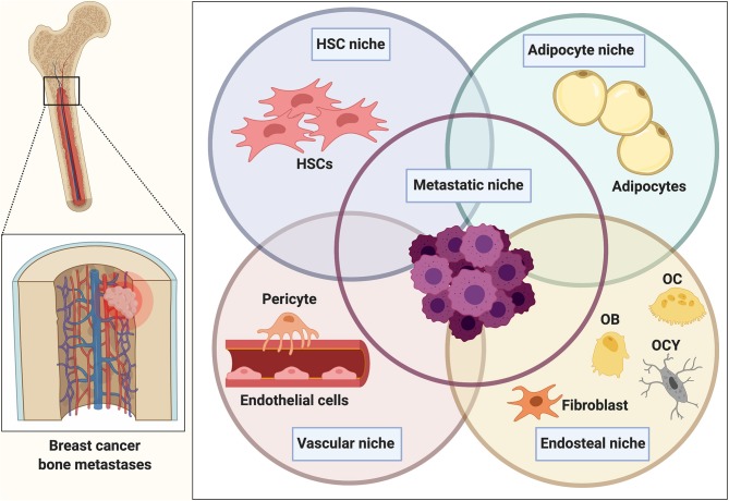
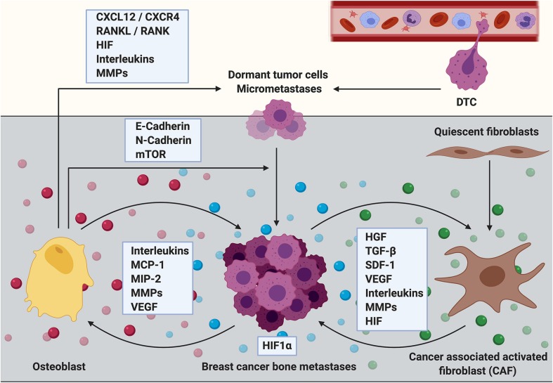
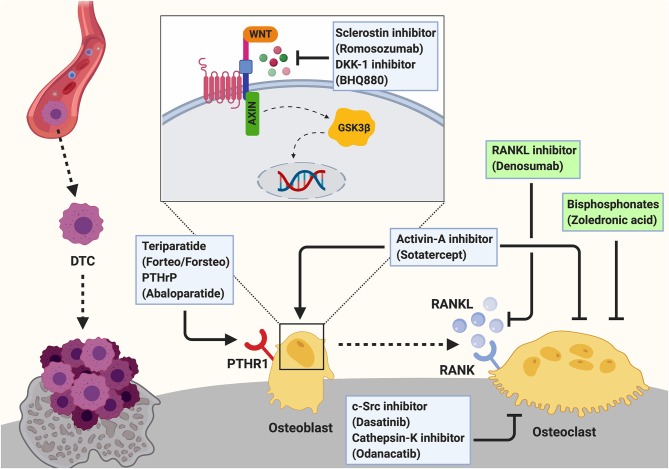
The establishment of bone metastasis remains one of the most frequent complications of patients suffering from advanced breast cancer. Patients with bone metastases experience high morbidity and mortality caused by excessive, tumor-induced and osteoclast-mediated bone resorption. Anti-resorptive treatments, such as bisphosphonates, are available to ease skeletal related events including pain, increased fracture risk, and hypercalcemia. However, the disease remains incurable and 5-year survival rates for these patients are below 25%. Within the bone, disseminated breast cancer cells localize in "metastatic niches," special microenvironments that are thought to regulate cancer cell colonization and dormancy as well as tumor progression and subsequent development into overt metastases. Precise location and composition of this "metastatic niche" remain poorly defined. However, it is thought to include an "endosteal niche" that is composed of key bone cells that are derived from both, hematopoietic stem cells (osteoclasts), and mesenchymal stromal cells (osteoblasts, fibroblasts, adipocytes). Our knowledge of how osteoclasts drive the late stage of the disease is well-established. In contrast, much less is known about the interaction between osteogenic cells and disseminated tumor cells prior to the initiation of the osteolytic phase. Recent studies suggest that mesenchymal-derived cells, including osteoblasts and fibroblasts, play a key role during the early stages of breast cancer bone metastasis such as tumor cell homing, bone marrow colonization, and tumor cell dormancy. Hence, elucidating the interactions between breast cancer cells and mesenchymal-derived cells that drive metastasis progression could provide novel therapeutic approaches and targets to treat breast cancer bone metastasis. In this review we discuss evidences reporting the interaction between tumor cells and endosteal niche cells during the early stages of breast cancer bone metastasis, with a particular focus on mesenchymal-derived osteoblasts and fibroblasts.

Keywords: bone metastases; breast cancer; endosteal niche; fibroblast; microenvironment; osteoblast.


Figures

Figure 1
Figure 1
The bone metastatic niche. Once homed to bone, tumor cells are exposed to a heterogeneous microenvironment that is comprised of various individual cellular entities. The complex interplay between osteoblasts and osteoclasts during bone remodeling in addition to the presence of various other bone marrow-derived populations makes the bone microenvironment a favorable and supportive environment (metastatic niche) for disseminated cancer cells. Within bone, the metastatic niche is thought to be comprised of a hematopoietic stem cell niche (HSCs), endosteal (osteoclasts (OC), osteoblasts (OB), osteocytes (OCY), fibroblasts), and vascular niche (endothelial cells, pericytes). Several findings also implicate a role of the bone marrow adipocyte niche in bone metastasis. The interaction and overlap between the niches remain to be determined and resulted in the generalized term of the “metastatic niche” that is thought to regulate homing, survival and dormancy of tumor cells.
Figure 2
Figure 2
The role of osteoblasts and cancer associated fibroblasts during the establishment and progression of breast cancer bone metastasis. Cells of mesenchymal origin including osteoblasts and cancer associated fibroblasts (CAFs) are increasingly recognized to contribute to the establishment and progression of breast cancer bone metastasis. Osteoblasts express cytokines including C-X-C motif chemokine ligand 12 (CXCL12, also referred to as stromal derived factor 1, SDF-1) and receptor activator of NF-κB ligand (RANKL) that promote breast cancer cell dissemination and metastatic growth through interaction with their corresponding receptors (CXCR4 and RANK, respectively) that are expressed by the cancer cells. Breast cancer micrometastases have also been shown to be surrounded by osteoblastic cells. The interaction between breast cancer cells and osteoblasts could partially be mediated via heterotypic adherens junctions using E-, and N-cadherins resulting in an enhanced mTOR activity in cancer cells and consequently in the transition from dormant tumor cells into overt metastases. Osteoblasts also express high levels of extracellular matrix remodeling proteins (MMPs) in addition to reduced presence of inflammatory cytokines such as interleukins (ILs) upon cancer cell stimulation. Thereby osteoblasts could regulate breast cancer cell dormancy in the bone microenvironment. In contrast, metastatic breast cancer cells increase the production of inflammatory cytokines such as IL-6 and IL-8, monocyte chemoattractant protein−1 (MCP-1), macrophage—inflammatory protein 2 (MIP-2) and vascular endothelial growth factor (VEGF) in osteoblasts, thereby promoting breast cancer cell invasiveness and metastasis progression. Although usually quiescent in normal tissue, fibroblasts acquire an activated phenotype during processes such as wound healing or inflammation. Activated fibroblasts in the tumor stroma are called cancer associated fibroblasts (CAFs). They produce growth factors that contribute to disease progression including hepatocyte growth factor (HGF), transforming growth factor beta (TGF-β), stromal derived factor 1 (SDF-1 or CXCL12), VEGF, IL-6, and other ILs in addition to MMPs. All of these factors promote primary tumor growth and it can be hypothesized that CAFs could similarly mediate the growth of breast cancer bone metastases. CAFs are known to induce extracellular matrix remodeling and alter the stiffness of tissues thereby facilitating tumor cell invasion, dissemination and/or metastasis. CAF-induced matrix remodeling and CAF invasion have been shown to be supported by hypoxia inducible factor−1 alpha (HIF1α). In turn, an increased expression of HIF1α might stimulate the tumor growth promoting function of CAFs.
Figure 3
Figure 3
Targeting the osteogenic niche to treat breast cancer bone metastasis. Breast cancer bone metastases remain incurable once osteolytic lesions have developed. Palliative treatment often includes the administration of osteoclast-targeted, anti-resorptive agents including bisphosphonates (e.g., Zoledronic acid) or the anti-RANKL antibody Denosumab to prevent the cancer-induced bone resorption. These two agents are the only approved treatments for cancer induced bone disease (indicated by green box). Additionally, c-Src (Dasatinib) and Cathepsin-K (Odanacatib) inhibitors are under investigation for the treatment of breast cancer bone metastasis. As these anti-resorptive agents are not able to restore the cancer-induced bone loss, augmenting osteoblast function by anabolic treatments has been proposed as a potential therapeutic approach and several agents are investigated experimentally and/or in clinical trials. Bone anabolic treatments including the administration of a recombinant fragment of PTH (Teriparatide; Forteo/Forsteo) or Parathyroid hormone related protein (PTHrP; Abaloparatide) are approved for the treatment of osteoporosis. However, these drugs cannot be prescribed for patients with bone metastases. Another bone anabolic agent Romosozumab, an antibody against the Wnt signaling inhibitor Sclerostin, increases bone formation and bone mass by activating the Wnt pathway in osteoblasts. Similarly, Dkk-1 inhibitors (e.g., BHQ880) allow active Wnt signaling in osteoblasts thereby increasing osteoblast activity. Inhibition of Activin-A signaling has been shown to prevent cancer-induced bone destruction. Additionally, Activin-A inhibitors (e.g., Sotatercept) have been shown to stimulate osteoblastogenesis while decreasing osteoclast activity to promote bone formation. Hence, they could potentially be a novel approach for the treatment of cancer induced bone disease.

References

    1. Weilbaecher KN, Guise TA, McCauley LK. Cancer to bone: a fatal attraction. Nat Rev Cancer. (2011) 11:411–25. 10.1038/nrc3055 - DOI - PMC - PubMed
    1. Paget S. The distribution of secondary growths in cancer of the breast. 1889. Cancer Metastasis Rev. (1989) 8:98–101. - PubMed
    1. Croucher PI, McDonald MM, Martin TJ. Bone metastasis: the importance of the neighbourhood. Nat Rev Cancer. (2016) 16:373–86. 10.1038/nrc.2016.44 - DOI - PubMed
    1. Mundy GR. Mechanisms of bone metastasis. Cancer. (1997) 80(Suppl. 8):1546–56. 10.1002/(SICI)1097-0142(19971015)80:8+<1546::AID-CNCR4>3.0.CO;2-I - DOI - PubMed
    1. Clines GA, Guise TA. Molecular mechanisms and treatment of bone metastasis. Expert Rev Mol Med. (2008) 10:e7. 10.1017/S1462399408000616 - DOI - PubMed

LinkOut - more resources