Review
. 2019 Aug 20:5:14.
doi: 10.1186/s42234-019-0030-2. eCollection 2019.

Neural activity regulates autoimmune diseases through the gateway reflex

Affiliations
Review

Neural activity regulates autoimmune diseases through the gateway reflex

Andrea Stofkova et al. Bioelectron Med. .

Abstract

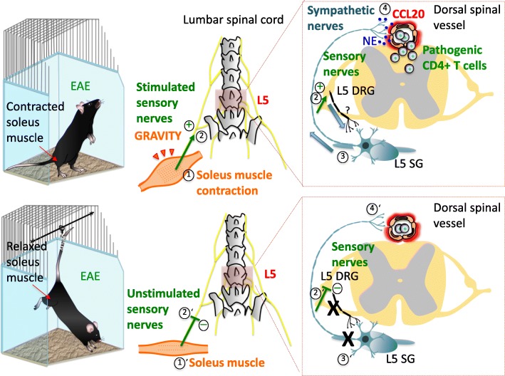
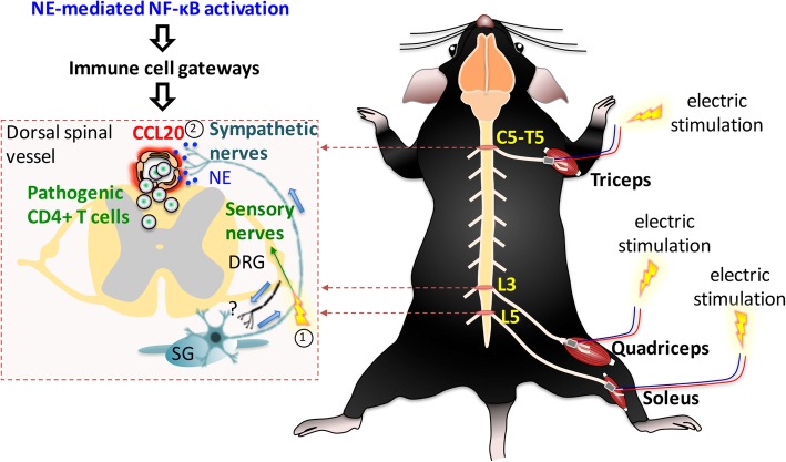
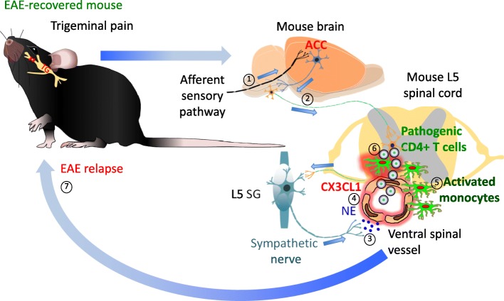
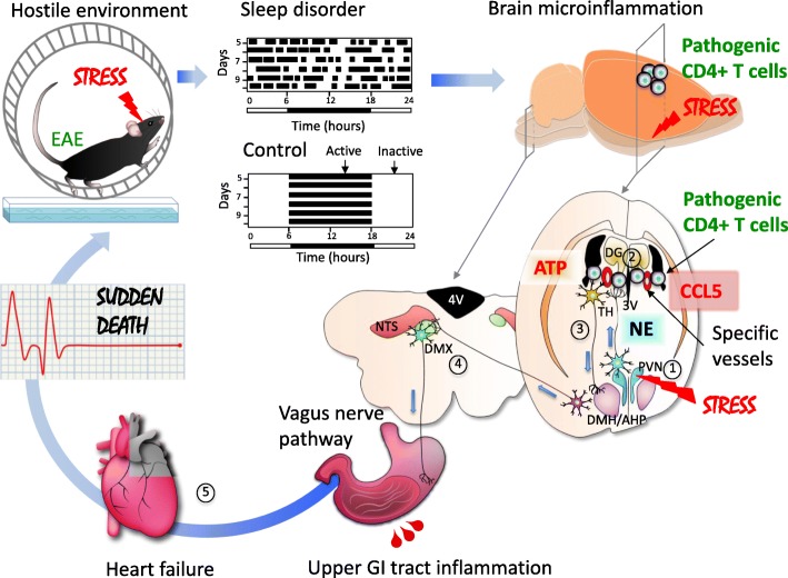
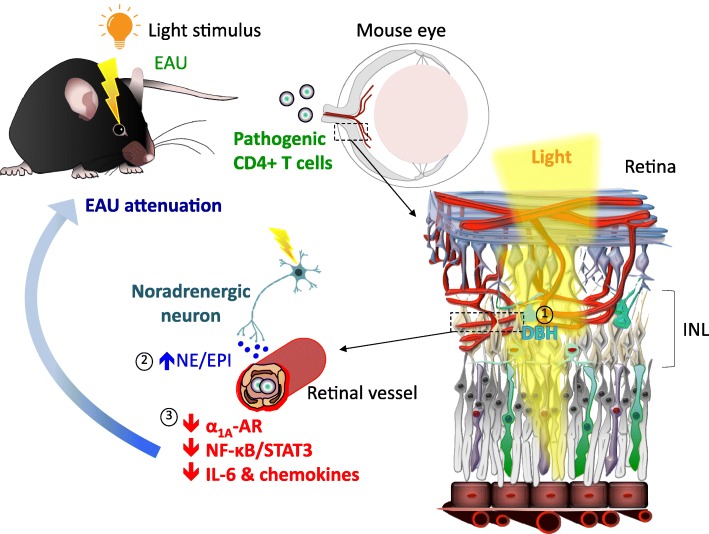
The brain, spinal cord and retina are protected from blood-borne compounds by the blood-brain barrier (BBB), blood-spinal cord barrier (BSCB) and blood-retina barrier (BRB) respectively, which create a physical interface that tightly controls molecular and cellular transport. The mechanical and functional integrity of these unique structures between blood vessels and nervous tissues is critical for maintaining organ homeostasis. To preserve the stability of these barriers, interplay between constituent barrier cells, such as vascular endothelial cells, pericytes, glial cells and neurons, is required. When any of these cells are defective, the barrier can fail, allowing blood-borne compounds to encroach neural tissues and cause neuropathologies. Autoimmune diseases of the central nervous system (CNS) and retina are characterized by barrier disruption and the infiltration of activated immune cells. Here we review our recent findings on the role of neural activity in the regulation of these barriers at the vascular endothelial cell level in the promotion of or protection against the development of autoimmune diseases. We suggest nervous system reflexes, which we named gateway reflexes, are fundamentally involved in these diseases. Although their reflex arcs are not completely understood, we identified the activation of specific sensory neurons or receptor cells to which barrier endothelial cells respond as effectors that regulate gateways for immune cells to enter the nervous tissue. We explain this novel mechanism and describe its role in neuroinflammatory conditions, including models of multiple sclerosis and posterior autoimmune uveitis.


Conflict of interest statement

Competing interestsThe authors declare that they have no competing interests.

Figures

Fig. 1
Fig. 1
Gravity gateway reflex. In free-moving mouse (upper panel) with adoptive-transfer EAE, pathogenic CD4+ T cells infiltrate the spinal cord parenchyma at the fifth lumbar (L5) level. This process is triggered by soleus muscle contraction (1) that counteracts gravity to maintain stability and body posture. Stimulated sensory nerves arising from the soleus muscle (2) and entering the L5 spinal cord through dorsal root stimulate sympathetic nerves (3) at the L5 spinal cord level via an unidentified neural circuit (?). This leads to chemokine expression including CCL20 in regional dorsal vessels of the spinal cord (4), establishing an immune cell gateway for migration of pathogenic CD4+ T cells across the BSCB. In tail-suspended mouse (lower panel) with adoptive-transfer EAE, soleus muscle relief from gravitational force (1’) prevents regional activation of sensory- (2’) and sympathetic-nerves (3’), and infiltration of pathogenic CD4+ T cells in the L5 spinal cord (4’). Abbreviations: EAE, experimental autoimmune encephalomyelitis; DRG, dorsal root ganglion; SG, sympathetic ganglion; NE, norepinephrine; BSCB, blood-spinal cord barrier
Fig. 2
Fig. 2
Electronic gateway reflex. Artificial neural activations by weak electric stimulations can induce the gateway reflex at individual blood vessels in the CNS. Electric stimulations to the triceps (1) induce chemokine upregulation at the dorsal vessels of the fifth cervical (C5) to fifth thoracic (T5) spinal cord (2) via the inflammation amplifier, which is mediated by NE secretion. Likewise, electric stimulations to the quadriceps (1) trigger chemokine upregulation at the L3 dorsal vessels (2), whereas the L5 gateway is formed by electric stimulation to the soleus muscles. Abbreviations: DRG, dorsal root ganglion; SG, sympathetic ganglion; NE, norepinephrine
Fig. 3
Fig. 3
Pain gateway reflex. In a transfer EAE-recovered mouse, induction of trigeminal pain by ligation of the infraorbital sensory nerve causes EAE relapse. In this condition, pathway carrying nociceptive information activates the ACC (1), a pain-processing area of the brain, which in turn stimulates sympathetic centers (2) and their neural output to the ventral spinal vessels (3). NE release around these vessels induces chemokine CX3CL1 expression from activated monocytes and endothelial cells (4). CX3CL1 recruits these activated monocytes around the ventral vessels in an autocrine/paracrine manner (5). Because the L5 spinal cord contains a high number of activated monocytes in EAE-recovered mice, the L5 region is most affected in terms of the accumulation of activated monocytes. Activated monocytes present MOG antigens to reactivate circulating pathogenic CD4+ T cells (6), thus causing their accumulation in the spinal cord and EAE relapse (7). Abbreviations: EAE, experimental autoimmune encephalomyelitis; ACC, anterior cingulate cortex; NE, norepinephrine; MOG, myelin oligodendrocyte glycoprotein; TG, trigeminal ganglion
Fig. 4
Fig. 4
Stress gateway reflex. Hostile environment induced by a specific cage with water and a free rotation wheel causes chronic stress associated with sleep disorder. In a mouse with adoptive-transfer EAE, this chronic stress stimulates noradrenergic neurons in the PVN (1), which then project to bilateral specific vessels surrounded by the third ventricle (3V), dentate gyrus (DG) and thalamus (TH) (2) to upregulate chemokines including CCL5 and recruit pathogenic CD4+ T cells plus MHC class II+ monocytes. The resulted microinflammation activates a neural pathway involving the DMH/AHP and DMX via ATP production (3). Hyperactivation of the vagus pathway particularly distributed the upper gastrointestinal (GI) tract causes a severe damage in the upper GI tract followed by a heart failure with sudden death (5). Abbreviations: AHP, anterior hypothalamic area; EAE, experimental autoimmune encephalomyelitis; PVN, paraventricular nucleus of the hypothalamus; DMH, dorsomedial nucleus of the hypothalamus; DMX, dorsal motor nucleus of the vagus nerve; NTS, nucleus tractus solitarii; 4V, fourth ventricle
Fig. 5
Fig. 5
Light gateway reflex. Exposure to photopic light in actively induced EAU attenuates retinal inflammation. This process is mediated through light-induced expression of DBH, an enzyme essential for the synthesis of NE and EPI, by retinal neurons located in the INL (1). The increased NE and EPI levels down-regulate retinal α1AAR expression, which is followed by reduced NF-κB and STAT3 activation and decreased chemokine and IL-6 expressions in the retina (2)(3). This protects the BRB integrity and suppresses recruitment of immune cells including IRBP-specific pathogenic CD4+ T cells in the retina. Abbreviations: EAU, experimental autoimmune uveoretinitis; DBH, dopamine beta-hydroxylase; INL, inner nuclear layer; NE, norepinephrine, EPI: epinephrine; α1AAR, alpha-1A adrenoceptor; BRB, blood-retina barrier; IRBP, interphotoreceptor retinoid-binding protein

References

    1. Abbott NJ, Rönnbäck L, Hansson E. Astrocyte-endothelial interactions at the blood-brain barrier. Nat Rev Neurosci. 2006;7(1):41–53. doi: 10.1038/nrn1824. - DOI - PubMed
    1. Akinaga J, Lima V, Kiguti LR, Hebeler-Barbosa F, Alcántara-Hernández R, García-Sáinz JA, Pupo AS. Differential phosphorylation, desensitization, and internalization of α1A-adrenoceptors activated by norepinephrine and oxymetazoline. Mol Pharmacol. 2013;83(4):870–881. doi: 10.1124/mol.112.082313. - DOI - PubMed
    1. Andersson U, Tracey KJ. Reflex principles of immunological homeostasis. Annu Rev Immunol. 2012;30:313–335. doi: 10.1146/annurev-immunol-020711-075015. - DOI - PMC - PubMed
    1. Arima Y, Harada M, Kamimura D, Park JH, Kawano F, Yull FE, Kawamoto T, Iwakura Y, Betz UA, Márquez G, Blackwell TS, Ohira Y, Hirano T, Murakami M. Regional neural activation defines a gateway for autoreactive T cells to cross the blood-brain barrier. Cell. 2012;148(3):447–457. doi: 10.1016/j.cell.2012.01.022. - DOI - PubMed
    1. Arima Y, Kamimura D, Atsumi T, Harada M, Kawamoto T, Nishikawa N, Stofkova A, Ohki T, Higuchi K, Morimoto Y, Wieghofer P, Okada Y, Mori Y, Sakoda S, Saika S, Yoshioka Y, Komuro I, Yamashita T, Hirano T, Prinz M, Murakami M. A pain-mediated neural signal induces relapse in murine autoimmune encephalomyelitis, a multiple sclerosis model. Elife. 2015;11:4. doi: 10.7554/eLife.08733. - DOI - PMC - PubMed

LinkOut - more resources