Review
. 2020 Apr;42(2):131-142.
doi: 10.1007/s00281-020-00792-2. Epub 2020 Mar 30.

Epigenetic lifestyle of Epstein-Barr virus

Affiliations
Review

Epigenetic lifestyle of Epstein-Barr virus

Alexander Buschle et al. Semin Immunopathol. 2020 Apr.

Abstract

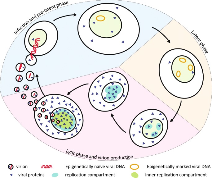
Epstein-Barr virus (EBV) is a model of herpesvirus latency and epigenetic changes. The virus preferentially infects human B-lymphocytes (and also other cell types) but does not turn them straight into virus factories. Instead, it establishes a strictly latent infection in them and concomitantly induces the activation and proliferation of infected B cells. How the virus establishes latency in its target cells is only partially understood, but its latent state has been studied intensively by many. During latency, several copies of the viral genome are maintained as minichromosomes in the nucleus. In latently infected cells, most viral genes are epigenetically repressed by cellular chromatin constituents and DNA methylation, but certain EBV genes are spared and remain expressed to support the latent state of the virus in its host cell. Latency is not a dead end, but the virus can escape from this state and reactivate. Reactivation is a coordinated process that requires the removal of repressive chromatin components and a gain in accessibility for viral and cellular factors and machines to support the entire transcriptional program of EBV's ensuing lytic phase. We have a detailed picture of the initiating events of EBV's lytic phase, which are orchestrated by a single viral protein - BZLF1. Its induced expression can lead to the expression of all lytic viral proteins, but initially it fosters the non-licensed amplification of viral DNA that is incorporated into preformed capsids. In the virions, the viral DNA is free of histones and lacks methylated cytosine residues which are lost during lytic DNA amplification. This review provides an overview of EBV's dynamic epigenetic changes, which are an integral part of its ingenious lifestyle in human host cells.

Keywords: Chromatin; Herpesvirus; Infection; Latency; Reactivation.


Conflict of interest statement

The authors declare that they have no conflict of interest.

Figures

Fig. 1
Fig. 1
The tripartite life cycle of EBV. Infection and pre-latent phase (light blue segment). Upon infection, a virion releases it epigenetically naïve linear DNA (red) into its host cell. The viral DNA circularizes and makes its way into the nucleus (light green). As the incoming DNA is not epigenetically repressed, many viral proteins (blue triangles) are expressed at low levels initially. During this phase, the host cell grows in size and starts to proliferate (not shown). After a couple of days, histones and nucleosomes are positioned, and mostly repressive epigenetic marks are established on the viral, plasmid-like minichromosome. Latent phase (light yellow segment). EBV expresses only few latent EBV proteins to maintain and preserve its epigenetically repressed DNA in latently infected cells, and EBV DNA acquires a very high level of CpG methylation (not shown). Additionally, this strategy avoids the detection of viral antigens by the immune system. In the nucleus, several copies of viral repressed EBV minichromosomes accumulate via an unknown mechanism. Latent phase and virion production (light red segment). Upon reactivation of the virus’ lytic phase, EBV starts to express lytic viral proteins, repressed viral chromatin opens up, and replication compartments (turquoise ovals) start to form. The full cascade of viral proteins is synthesized eventually, which counteracts the immune response of the host organism, supports lytic amplification of viral DNA, and provides structural proteins such as capsid components, tegument proteins, and glycoproteins. Replication compartments form, grow in dimension, and fuse during the massive replication of viral DNA. The newly synthesized viral DNA is packaged into viral capsid structures within the inner replication compartment (green area within the replication compartment). The newly replicated viral DNA is free of histones and lacks methylated CpG dinucleotides such that assembled virions contain epigenetically naïve viral DNA ready to infect new target cells.

References

    1. Adamson AL, Kenney S. The Epstein-Barr virus BZLF1 protein interacts physically and functionally with the histone acetylase CREB-binding protein. J Virol. 1999;73:6551–6558. doi: 10.1128/JVI.73.8.6551-6558.1999. - DOI - PMC - PubMed
    1. Albright ER, Kalejta RF. Canonical and variant forms of histone H3 are deposited onto the human cytomegalovirus genome during lytic and latent infections. J Virol. 2016;90:10309–10320. doi: 10.1128/JVI.01220-16. - DOI - PMC - PubMed
    1. Amon W, Binne UK, Bryant H, Jenkins PJ, Karstegl CE, Farrell PJ. Lytic cycle gene regulation of Epstein-Barr virus. J Virol. 2004;78:13460–13469. doi: 10.1128/JVI.78.24.13460-13469.2004. - DOI - PMC - PubMed
    1. Amon W, Farrell PJ. Reactivation of Epstein-Barr virus from latency. Rev Med Virol. 2005;15:149–156. doi: 10.1002/rmv.456. - DOI - PubMed
    1. Anonymous . Epstein-Barr virus; IARC monographs on the evaluation of carcinogenic risks to humans. Lyon: IARC; 1997. p. 1. - PMC - PubMed

Publication types

LinkOut - more resources