. 2020 May;19(5):e13107.
doi: 10.1111/acel.13107. Epub 2020 Mar 31.

Muscle atrophy-related myotube-derived exosomal microRNA in neuronal dysfunction: Targeting both coding and long noncoding RNAs

Affiliations

Muscle atrophy-related myotube-derived exosomal microRNA in neuronal dysfunction: Targeting both coding and long noncoding RNAs

Chia-Pei Yang et al. Aging Cell. 2020 May.

Abstract

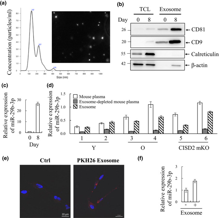
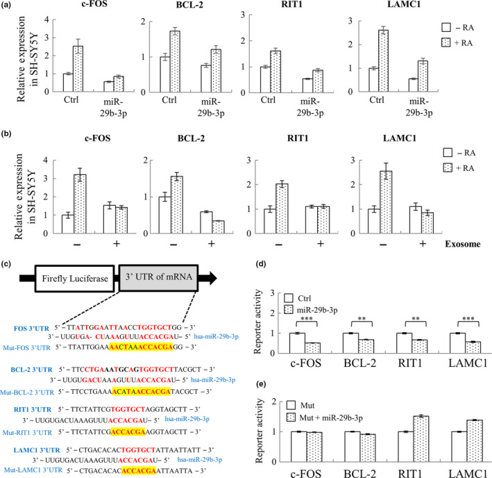
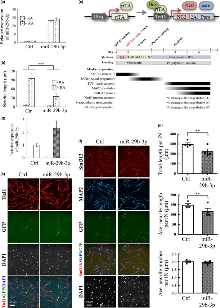
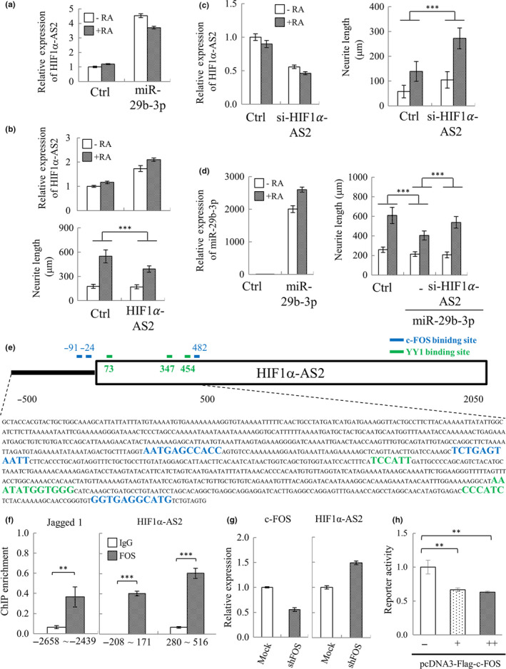
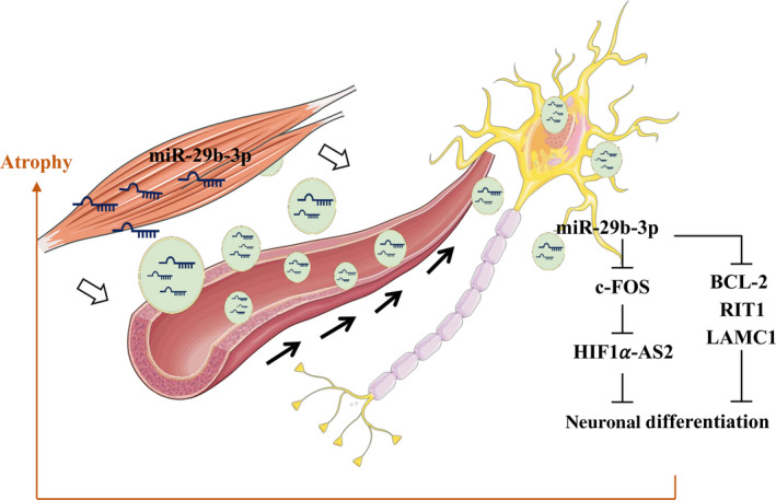
In mammals, microRNAs can be actively secreted from cells to blood. miR-29b-3p has been shown to play a pivotal role in muscle atrophy, but its role in intercellular communication is largely unknown. Here, we showed that miR-29b-3p was upregulated in normal and premature aging mouse muscle and plasma. miR-29b-3p was also upregulated in the blood of aging individuals, and circulating levels of miR-29b-3p were negatively correlated with relative appendicular skeletal muscle. Consistently, miR-29b-3p was observed in exosomes isolated from long-term differentiated atrophic C2C12 cells. When C2C12-derived miR-29b-3p-containing exosomes were uptaken by neuronal SH-SY5Y cells, increased miR-29b-3p levels in recipient cells were observed. Moreover, miR-29b-3p overexpression led to downregulation of neuronal-related genes and inhibition of neuronal differentiation. Interestingly, we identified HIF1α-AS2 as a novel c-FOS targeting lncRNA that is induced by miR-29b-3p through down-modulation of c-FOS and is required for miR-29b-3p-mediated neuronal differentiation inhibition. Our results suggest that atrophy-associated circulating miR-29b-3p may mediate distal communication between muscle cells and neurons.

Keywords: HIF-1α-AS2; aging; lncRNAs; miR-29b-3p; muscle atrophy.


Conflict of interest statement

The authors disclose no potential conflicts of interest.

Figures

Figure 1
Figure 1
Identification of miRNAs that are increased in atrophied muscle and in plasma of aged mice and humans. (a) Hematoxylin–eosin (HE) staining of quadriceps femoris muscles from 3‐month (Young, Y)‐ and 26‐month (Old, O)‐old wild‐type mice and 3‐month‐old CISD2 muscle‐specific knockout (mKO) mice revealed degeneration (black arrows) and cellular shrinkage (blue arrows) in the skeletal muscles of naturally aged and CISD2 mKO mice. (b) RT‐qPCR analysis of Atrogin‐1 and MuRF‐1 mRNA using femoris muscle RNA isolated from mice described in (a) (n = 6). (c) Venn diagram showing the number of the miRNAs up (left panel, red)‐ and down (right panel, blue)regulated in aged and CISD2 mKO muscle (RPKM > 1, fold change > 1.5x) (n = 3). (d) Hierarchical clustering of the 102 miRNAs shown in (c). Each column represents the miRNA expression in Y, O, and CISD2 mKO muscle. (e) Plasma RNA isolated from Y, O, and CISD2 mKO mice (n = 5) was subjected to RT‐qPCR. 9 miRNAs were detected in mouse plasma. (f) Plasma RNA isolated from young (21–30 years old) and old (71–80 years old) human subjects (n = 18) was subjected to RT‐qPCR. miR‐708‐3p and miR‐130b‐3p were significantly increased in plasma of elderly, and miRNA‐29b‐3p was increased with borderline statistical significance (p = .051). *p < .05, **p < .01, ***p < .001 by one‐way ANOVA
Figure 2
Figure 2
C2C12 myotube‐derived exosomes are taken up by RA‐differentiated SH‐SY5Y cells and results in increased level of miR‐29b‐3p. (a) Exosomes isolated by differential ultracentrifugation were analyzed using nanoparticle tracking analysis (NTA) and represented as size versus. concentration. (b) Immunoblotting assessing the exosome markers, CD81 and CD9, in C2C12 myotube‐derived exosomes enriched from undifferentiated (day 0) or 8 days differentiated cells. The exosome fraction is absent of endoplasmic reticulum (ER) marker calreticulin. β‐Actin was used as loading control. TCL, total cell lysate. (c) Exosomal RNA was purified by TRIzol, and miR‐29b‐3p quantity was determined by RT‐qPCR. (d) RNA from mouse plasma, exosome, and exosome‐depleted plasma was purified by TRIzol, and miR‐29b‐3p quantity was determined by RT‐qPCR. (e) SH‐SY5Y cells were induced for differentiation with 10 μM retinoic acid (RA) for 72 hr. RA‐differentiated SH‐SY5Y cells were maintained in the absence (left) or presence (right) of PKH26‐labeled exosomes (red). 24 hr after co‐incubation, cells were fixed, stained, and visualized by confocal (63x) microscope, demonstrating the presence of exosomes within cells. (f) RNA from RA‐differentiated SH‐SY5Y cells co‐cultured with or without long‐term differentiated C2C12 myotube‐derived exosomes was purified by TRIzol, and miR‐29b‐3p quantity was determined by RT‐qPCR. Error bars show mean ± SD (n = 3)
Figure 3
Figure 3
miR‐29b‐3p directly targets neuronal‐related genes c‐FOS, BCL‐2, RIT1, and LAMC1. (a) SH‐SY5Y cells were transiently transduced with lentivirus carrying control or pLenti4‐CMV/TO‐miR‐29b‐3p vector. 48 hr after transduction, SH‐SY5Y cells were treated with 10 μM RA for another 72 hr, followed by total RNA isolation and RT‐qPCR quantification of c‐FOS, BCL‐2, RIT1, and LAMC1. (b) SH‐SY5Y cells were treated with exosome for 24 hr and then induced for differentiation with 10 μM RA for 72 another hours. RNA from RA‐differentiated SH‐SY5Y cells co‐cultured with or without long‐term differentiated C2C12 myotube‐derived exosomes was purified, followed by quantification of c‐FOS, BCL‐2, RIT1, and LAMC1 using RT‐qPCR. (c) Structure of the luciferase reporter construct and the predicted miR‐29b‐3p binding site on the 3’UTR of c‐FOS, BCL‐2, RIT1, and LAMC1. (d and e) The luciferase reporter plasmids containing either miR‐29b‐3p binding site (d) or miR‐29b‐3p binding‐deficient mutant (Mut) (e) were co‐transfected with miR‐29b‐3p expression construct into 293T cells. Luciferase reporter assay results showing that c‐FOS, BCL‐2, RIT1, and LAMC1 were direct targets of miR‐29b‐3p. Error bars show mean ± SD (n = 3). **p < .01, ***p < .001 by Student's t test
Figure 4
Figure 4
miR‐29b‐3p inhibits SH‐SY5Y and iNs’ cell differentiation. (a) SH‐SY5Y cells were transiently transduced with lentivirus carrying control or pLenti4‐CMV/TO‐miR‐29b‐3p vector. 48 hr after transduction, SH‐SY5Y cells were treated with 10 μM RA for another 72 hr, followed by total RNA isolation and RT‐qPCR quantification of miR‐29b‐3p. (b) SH‐SY5Y cells treated as described in (a) were stained with cell membrane and nucleus and used for quantification of neurite length. (c) Vector design for NG2‐mediated conversion of hiPSCs into glutamatergic neurons (upper panel). Flow diagram depicting the workflow involved in the generation of glutamatergic neurons (middle panel). hiPSCs were sequentially infected with lentivirus‐expressing rtTA and NG2‐puromycin resistance fusion protein linked by T2A sequence. Following lentivirus transduction and Dox treatment, hiPSCs were selected by puromycin for 24 hr and then re‐seeded for analysis. Time course of biomarkers expression following iNs’ induction is shown (lower panel). (d) Total RNA isolation and RT‐qPCR quantification of miR‐29b‐3p on day 3. (e and f) Representative images of iNs on day 4. The iN cells infected with control and miR‐29b‐3p construct were fixed and immunostained with anti‐TuJ1 (e, red), anti‐MAP2 (f, blue), and anti‐Smi312 (f, red) antibodies. (g) Quantification of the total length (upper panel), average neurite length (middle panel), and average neurite number (lower panel) of TuJ1‐positive neurites in GFP‐positive cells. Data are presented as mean ± SEM (N = 4). ***p < .001 by two‐way ANOVA (b) and *p < .05, **p < .01 by Student's t test of (g)
Figure 5
Figure 5
Identification of HIF1α‐AS2 as a novel miR‐29b‐3p co‐upregulated lncRNA that negatively modulates RA‐induced SH‐SY5Y differentiation. (a) SH‐SY5Y cells were transfected with pre‐miR‐29b‐3p. 24 hr after transfection, cells were treated with 10 μM RA for another 72 hr. Total RNA purified from cells was subjected to lncRNA qPCR array analysis. Array result for HIF1α‐AS2 is shown. (b) SH‐SY5Y‐HIF1α‐AS2 and control cells were treated with 10 μM RA for 72 hr. The levels of HIF1α‐AS2 were measured in both RA‐treated and RA‐untreated cells by RT‐qPCR (upper panel). SH‐SY5Y cells were stained as described in Figure 4b, and neurite length was quantified (lower panel). (c) SH‐SY5Y cells were transiently transfected with siRNA specific for HIF1α‐AS2 (si‐HIF1α‐AS2). 24 hr after transfection, the cells were treated with 10 μM RA for another 72 hr. HIF1α‐AS2 levels (left panel) and neurite length (right panel) were quantified as described in (b). (d) SH‐SY5Y cells were co‐transfected with pre‐miR‐29b‐3p and si‐HIF1α‐AS2. 24 hr after transfection, cells were treated with 10 μM RA for 72 hr. The expression level of miR‐29b‐3p (left panel) and neurite length (right panel) was quantified as described in (b). (e) Schematic representation of the putative c‐FOS binding sites in HIF1α‐AS2 promoter (TSS ± 500 bp) predicted by JASPAR database. (f) ChIP‐qPCR analysis using anti‐c‐FOS‐specific antibody revealed direct binding of c‐FOS to the promoter region of HIF1α‐AS2 (right panel). Jagged 1 promoter is the positive control of ChIP (left panel). (g) SH‐SY5Y cells were transiently transduced with lentivirus carrying control or pLKO.1‐shFOS (TRCN0000016004) vector. 72 hr after transduction, knockdown of c‐FOS (left panel) and expression of HIF1α‐AS2 (right panel) were detected by RT‐qPCR. (h) The luciferase reporter plasmid containing HIF1α‐AS2 promoter was co‐transfected with pcDNA3‐Flag‐c‐FOS into 293T cells. Luciferase reporter assay results showing that c‐FOS directly targets the HIF1α‐AS2 promoter. Error bars show mean ± SD (n = 3). **p < .01, ***p < .001 by Student's t test (f), one‐way ANOVA (h), and two‐way ANOVA (b, c and d)
Figure 6
Figure 6
A schematic model of myotube‐derived exosomal miR‐29b‐3p in modulating neuronal cell function. miR‐29b‐3p‐containing exosomes released from atrophied muscle can be transported via the circulation and transferred to neuronal cells. Increased miR‐29b‐3p levels in recipient cells lead to (i) downregulation of neuronal differentiation‐related genes BCL‐2, RIT1, and LAMC1, and (ii) downregulation of c‐FOS, de‐repression of HIF1α‐AS2, and consequently inhibition of neuronal differentiation. The model depicts how atrophy‐associated exosomal miR‐29b‐3p may mediate distal communication between muscle and neuronal cells

References

    1. Akhtar, R. S. , Ness, J. M. , & Roth, K. A. (2004). Bcl‐2 family regulation of neuronal development and neurodegeneration. Biochimica Et Biophysica Acta, 1644, 189–203. 10.1016/j.bbamcr.2003.10.013 - DOI - PubMed
    1. Arroyo, J. D. , Chevillet, J. R. , Kroh, E. M. , Ruf, I. K. , Pritchard, C. C. , Gibson, D. F. , … Tewari, M. (2011). Argonaute2 complexes carry a population of circulating microRNAs independent of vesicles in human plasma. Proceedings of the National Academy of Sciences, 108, 5003–5008. 10.1073/pnas.1019055108 - DOI - PMC - PubMed
    1. Bains, W. , Ponte, P. , Blau, H. , & Kedes, L. (1984). Cardiac actin is the major actin gene product in skeletal muscle cell differentiation in vitro. Molecular and Cellular Biology, 4, 1449–1453. 10.1128/MCB.4.8.1449 - DOI - PMC - PubMed
    1. Bhome, R. , Goh, R. , Pickard, K. , Mellone, M. , Sayan, A. E. , & Mirnezami, A. (2017). Profiling the MicroRNA payload of exosomes derived from ex vivo primary colorectal fibroblasts. Methods in Molecular Biology, 1509, 115–122. - PMC - PubMed
    1. Bodine, S. C. , Latres, E. , Baumhueter, S. , Lai, V. K. , Nunez, L. , Clarke, B. A. , … Glass, D. J. (2001). Identification of ubiquitin ligases required for skeletal muscle atrophy. Science, 294, 1704–1708. 10.1126/science.1065874 - DOI - PubMed

Publication types