Review
. 2021 Jun 1;145(6):664-677.
doi: 10.5858/arpa.2019-0665-RA.

What is New in the 2019 World Health Organization (WHO) Classification of Tumors of the Digestive System: Review of Selected Updates on Neuroendocrine Neoplasms, Appendiceal Tumors, and Molecular Testing

Affiliations
Review

What is New in the 2019 World Health Organization (WHO) Classification of Tumors of the Digestive System: Review of Selected Updates on Neuroendocrine Neoplasms, Appendiceal Tumors, and Molecular Testing

Naziheh Assarzadegan et al. Arch Pathol Lab Med. .

Abstract

Context.—: The 5th edition of the World Health Organization classification of digestive system tumors discusses several advancements and developments in understanding the etiology, pathogenesis, and diagnosis of several digestive tract tumors.

Objective.—: To provide a summary of the updates with a focus on neuroendocrine neoplasms, appendiceal tumors, and the molecular advances in tumors of the digestive system.

Data sources.—: English literature and personal experiences.

Conclusions.—: Some of the particularly important updates in the 5th edition are the alterations made in the classification of neuroendocrine neoplasms, understanding of pathogenesis of appendiceal tumors and their precursor lesions, and the expanded role of molecular pathology in establishing an accurate diagnosis or predicting prognosis and response to treatment.


Figures

Figure 1.
Figure 1.
Ileal neuroendocrine tumor, grade 1. A and b, Low and medium power view; the tumor displays architectural patterns of cords, ribbons, and nests. C and D, Higher-power view of (A) shows nests of neoplastic cells with round to oval nuclei containing coarsely clumped, “salt and pepper” chromatin (hematoxylin-eosin, original magnifications ×2 [A] and ×10 [B]; original magnification ×100 [C and D]).
Figure 2.
Figure 2.
Small cell neuroendocrine carcinoma (SCNEC). A and B, SCNEC of the esophagus demonstrating solid sheets of small cells with high N:C ratio, fusiform nuclei, nuclear molding and scant cytoplasm arising in a background of high-grade dysplasia in Barrett esophagus. C, Immunohistochemical staining for synaptophysin shows diffuse staining with focal “dot-like” positivity in some cells. D, Ki-67 immunostaining, with a Ki-67 proliferation index of 90% (hematoxylin-eosin, original magnifications ×2 [A] and ×100 [B]; original magnifications ×200 [C] and ×100 [D]).
Figure 3.
Figure 3.
Large cell neuroendocrine carcinoma (LCNEC). A and B, LCNEC composed of sheets of large cells with dispersed chromatin, prominent nucleoli, and moderate-to-abundant eosinophilic cytoplasm (hematoxylin-eosin, original magnifications ×10 [A] and ×200 [B]).
Figure 4.
Figure 4.
Mixed neuroendocrine-non-neuroendocrine neoplasms (MiNEN). A, Low-power view; MiNEN of the gastric mucosa composed of adenocarcinoma and grade 1 neuroendocrine tumor (NET). B, Higher-power view of (A) shows the infiltrative glands of adenocarcinoma adjacent to grade 1 NET. C, Background of atrophic gastritis showing atrophic oxyntic glands and intestinal metaplasia. D, Immunohistochemical staining for synaptophysin shows diffuse and strong immunoreactivity in the neuroendocrine component whereas it is negative in the adenocarcinoma (hematoxylin-eosin, original magnifications ×10 [A] and ×100 [B and C]; original magnification ×200 [D]).
Figure 5.
Figure 5.
Appendiceal sessile serrated lesion without dysplasia. A, Low-power view; the crypts are elongated with serrated appearance. B, High-power view shows the dilatation and lateral spread of the crypts at the base. The muscularis mucosae is intact (hematoxylin-eosin, original magnifications ×10 [A] and ×100 [B]).
Figure 6.
Figure 6.
Low-grade appendiceal mucinous neoplasm. A, The mucosa is replaced by hypermucinous undulating epithelium. B, Higher-power view shows the epithelium lined by tall cells with low-grade cytological atypia and hyalinized, fibrotic submucosa with no significant inflammatory response (hematoxylin-eosin, original magnifications ×100 [A] and ×200 [B]).
Figure 7.
Figure 7.
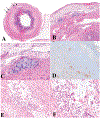
Goblet cell adenocarcinoma, low grade (A–D). A, Low-power view; the tumor infiltrates the muscularis propria of the appendix as indicated by the arrows. B and C, High-power view of (A) shows clusters of cohesive goblet-like cells. D, Immunohistochemical stain for synaptophysin (not required for diagnosis) shows strong immunoreactivity in this case. Goblet cell adenocarcinoma, high grade (E, F). E, Low-power view; the tumor is infiltrative and shows complex anastomosing tubules. F, High-power view highlights the complex architectures with streaming of discohesive tumor cells with high-grade cytological features, in contrast to cohesive clusters of low-grade lesions (hematoxylin-eosin, original magnifications ×2 [A], ×100 [B, E], ×200 [C, F]; original magnification ×100 [D]).

Comment in

References

    1. Basturk O, Yang Z, Tang LH, et al. The high-grade (WHO G3) pancreatic neuroendocrine tumor category is morphologically and biologically heterogenous and includes both well differentiated and poorly differentiated neoplasms. Am J Surg Pathol. 2015;39(5):683–690. - PMC - PubMed
    1. Nagtegaal ID, Odze RD, Klimstra D, et al.; for the WHO Classification of Tumours Editorial Board. The 2019 WHO classification of tumours of the digestive system. Histopathology. 2020;76(2):182–188. - PMC - PubMed
    1. Kim JY, Hong S. Recent updates on neuroendocrine tumors from the gastrointestinal and pancreatobiliary tracts. Arch Pathol Lab Med. 2016;140(5):437–448. - PubMed
    1. Rindi G, Klimstra DS, Abedi-Ardekani B, et al. A common classification framework for neuroendocrine neoplasms: an international agency for research on cancer (IARC) and world health organization (WHO) expert consensus proposal. Mod Pathol. 2018;31(12):1770–1786. - PMC - PubMed
    1. Lokuhetty D, White V, Watanabe R, Cree I. WHO classification of tumours of the digestive system. Lyon: International Agency for Research on Cancer; 2018.

MeSH terms