. 2020 Apr 1:9:e51576.
doi: 10.7554/eLife.51576.

Functionally heterogeneous human satellite cells identified by single cell RNA sequencing

Affiliations

Functionally heterogeneous human satellite cells identified by single cell RNA sequencing

Emilie Barruet et al. Elife. .

Abstract

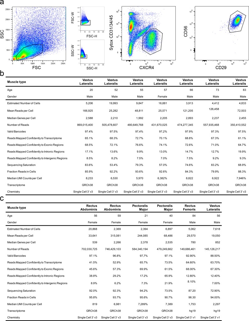
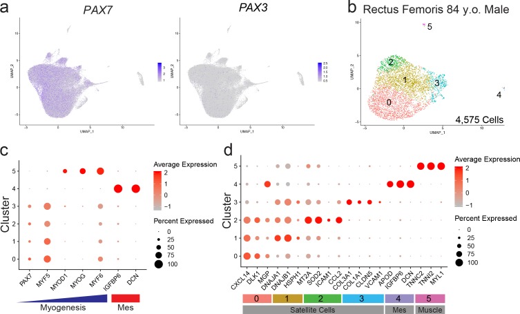
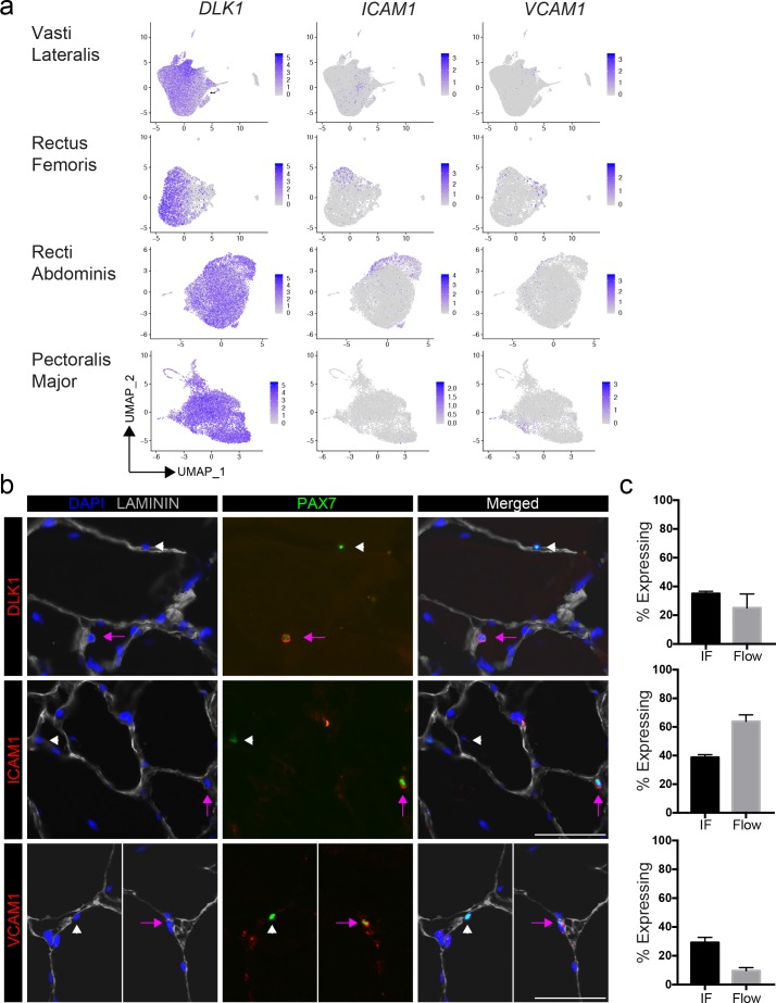
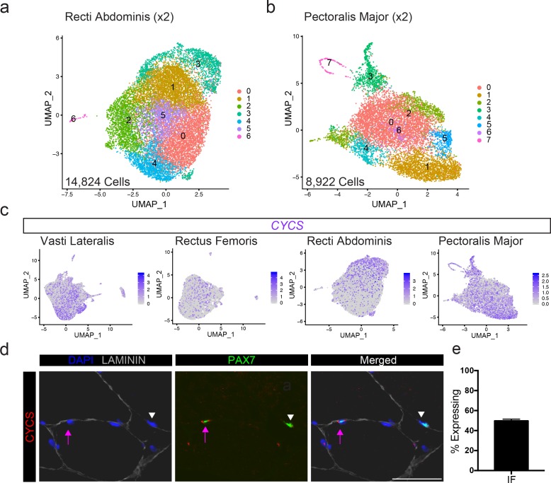
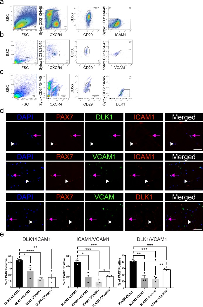
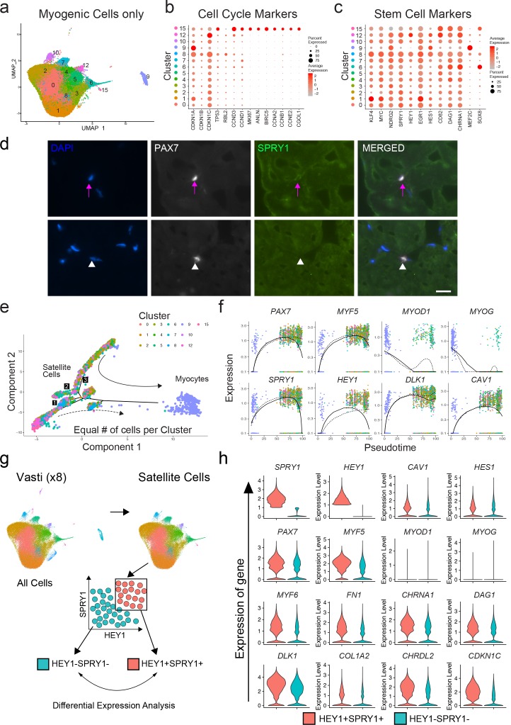
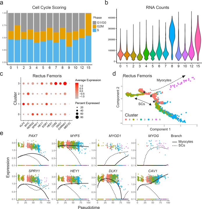
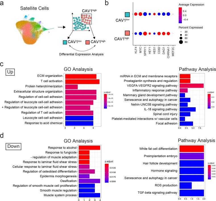
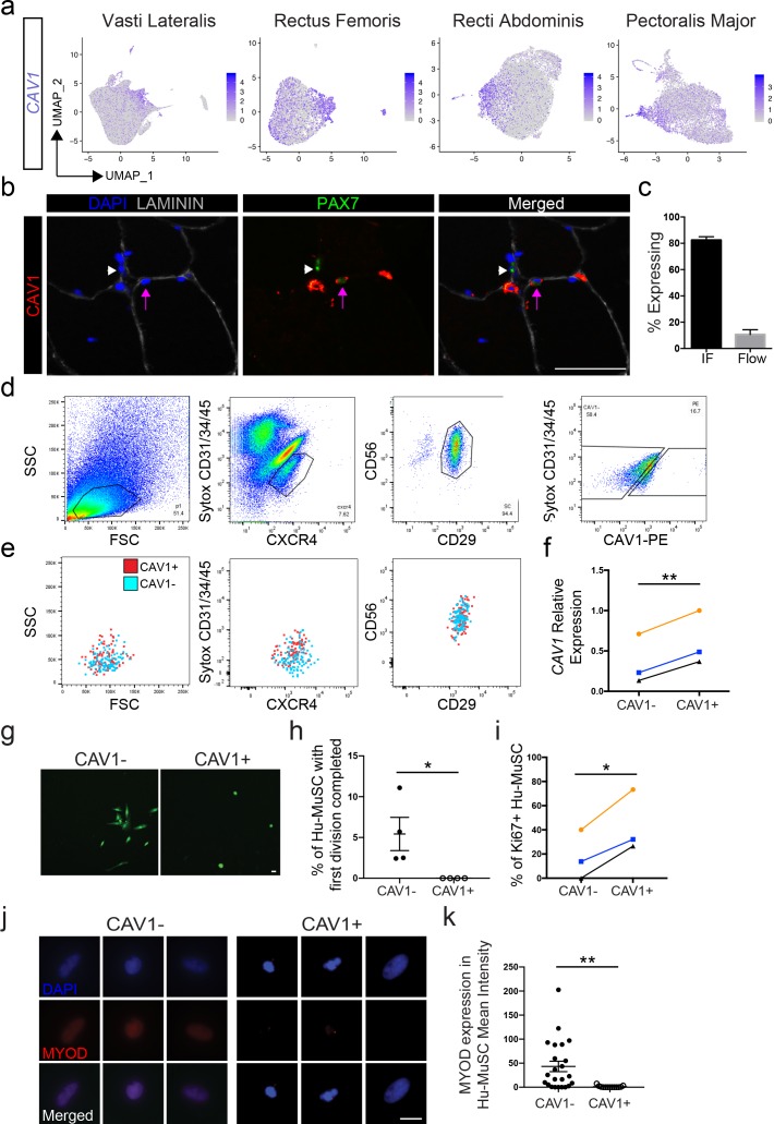
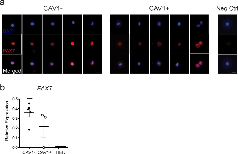
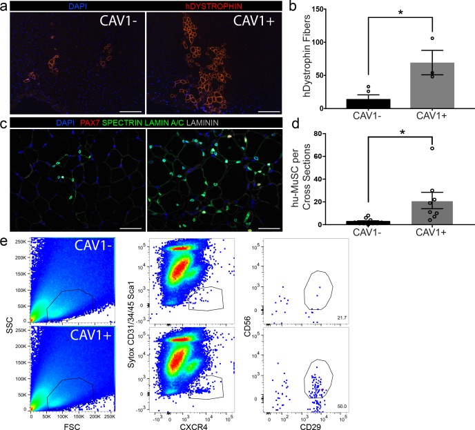
Although heterogeneity is recognized within the murine satellite cell pool, a comprehensive understanding of distinct subpopulations and their functional relevance in human satellite cells is lacking. We used a combination of single cell RNA sequencing and flow cytometry to identify, distinguish, and physically separate novel subpopulations of human PAX7+ satellite cells (Hu-MuSCs) from normal muscles. We found that, although relatively homogeneous compared to activated satellite cells and committed progenitors, the Hu-MuSC pool contains clusters of transcriptionally distinct cells with consistency across human individuals. New surface marker combinations were enriched in transcriptional subclusters, including a subpopulation of Hu-MuSCs marked by CXCR4/CD29/CD56/CAV1 (CAV1+). In vitro, CAV1+ Hu-MuSCs are morphologically distinct, and characterized by resistance to activation compared to CAV1- Hu-MuSCs. In vivo, CAV1+ Hu-MuSCs demonstrated increased engraftment after transplantation. Our findings provide a comprehensive transcriptional view of normal Hu-MuSCs and describe new heterogeneity, enabling separation of functionally distinct human satellite cell subpopulations.

Keywords: Human satellite cell transcriptome; human; human biology; medicine; muscle stem cell; regenerative medicine; satellite cell transplantation; stem cells.


Conflict of interest statement

EB, SG, KS, JW, SL, LB, AW, SX, ST, AB, JP No competing interests declared

Figures

Figure 1.
Figure 1.. Single cell RNA sequencing reveals heterogeneity of the human satellite cell transcriptome.
(a) Schematic diagram of the process of isolating human satellite cells from muscle biopsies, and then performing single cell RNA-seq to develop single cell transcriptomes leading to discovery of cell clusters, followed by in vivo validation. (b) UMAP plot of 68,108 cells isolated from eight vasti lateralis of male and female healthy subjects. Cells are clustered according to transcriptome similarity in 2D space. Each dot represents one cell, which are colored by cluster as identified by clustering analysis. (c) UMAP of each individual sample showing the distribution of cells in clusters for each sample. (d–f) Dot plots displaying expression of individual genes within each cluster. Each cluster is depicted on the y-axis and genes are labeled on the x-axis. Larger dot size represents more cells of that cluster expressing each gene; while color indicates the level of expression within those cells. (d) Dot plot displaying expression of genes associated with myogenesis, mesenchymal and hematopoietic markers in clusters 0–16. (e) Dot plot displaying the expression of top differentially expressed cluster markers in clusters 0–16. (f) ‘Road-UMAP’ with labeled cell types and key significantly regulated markers.
Figure 1—figure supplement 1.
Figure 1—figure supplement 1.. Flow cytometric sorting of CXCR4+/CD29+/CD56+ human satellite cells and quality control data from single cell RNA-sequencing experiments.
(a) Flow cytometry profiles from the isolation of CXCR4/CD29/CD56 human satellite cells from vastus lateralis muscle of a 55 year old male. (b–c) Quality control metrics for each of the single cell RNA-sequencing experiments of cells isolated from an 84 year old male (left) and a 56 year old male (right).
Figure 1—figure supplement 2.
Figure 1—figure supplement 2.. PAX7/3 expression in vasti lateralis and transcriptome from a rectus femoris muscle of human satellite cells.
(a) Feature map displaying the expression of PAX7 and PAX3 of satellite cells in eight pooled vasti lateralis human muscle. Of Note while the rectus femoris sample was prepared with the Chromium Single Cell 3' Reagent v1 kit, we found that the newer 3' Reagent v3 detected some genes including PAX7 with greater sensitivity. (b) UMAP of 5,062 cells isolated from the quadriceps muscle of a 84-year-old male. Cells are clustered according to transcriptome similarity in 2D space. Each dot represents one cell which are colored by cluster as identified by clustering analysis. (c) Dot plots displaying expression of individual genes within each cluster. Each cluster is depicted on the y-axis and genes are labeled on the x-axis. Larger dot size represents more cells of that cluster expressing each gene; while color indicates the over level of expression within those cells. (d) Dot plot displaying expression of genes associated with myogenesis and mesenchymal markers in clusters 0–5. (e) Dot plot displaying the expression of top differentially expressed cluster markers in clusters 0–5 similar to the eight vasti lateralis.
Figure 2.
Figure 2.. Validation of satellite cell clusters.
(a) Feature plots displaying localized gene expression for the genes DLK1, ICAM1, and VCAM1 within the 2D UMAP space of the eight combined vasti, the rectus femoris, the two combined rectus abdominis and the two combined pectoralis majors as shown in Figure 1b and Figure 1—figure supplement 2b, Figure 2—figure supplement 1a,b. Each dot represents a single cell. Deeper purple coloration represents increased expression. (b) Immunofluorescence staining of human satellite cells for validation markers within sections of human muscle. Pink arrows denote human satellite cells that are positive for the marker (scale, 50 µm). White arrowheads mark satellite cells that are negative for expression of the marker. (n = 3, biological replicates). (c) Bar plot displaying the quantification of DLK1, ICAM1, and VCAM1 expression of human satellite cells with both immunofluorescence staining and flow cytometry. (n = 3, biological replicates). Data presented as mean ± SEM.
Figure 2—figure supplement 1.
Figure 2—figure supplement 1.. Validation of satellite cell cluster marker CYCS across multiple human muscle type.
(a-b) UMAP plot of cells isolated from two recti abdominis and two pectoralis major. (c) Feature plot depicting localized gene expression of cluster marker CYCS in the UMAP 2D space in the eight vasti lateralis, rectus femoris, two recti abdominis and two pectoralis major. (d) Immunofluorescence staining of human satellite cells for CYCS within sections of human muscle (scale, 50 µm). Lavender arrows denote human satellite cells that are positive for CYCS. White arrowheads mark satellite cells that are negative for CYCS (n = 3, biological replicates). (e) Bar plot displaying the quantification CYCS expression of PAX7+ human satellite cells by immunofluorescence staining (n = 3, biological replicates). Data presented as mean ± SEM.
Figure 2—figure supplement 2.
Figure 2—figure supplement 2.. Heterogeneous expression of ICAM1, VCAM1 and DLK1 in human satellite cells.
(a-c) Representative flow cytometry profiles for CXCR4+/CD29+/CD56+ human satellite cells expressing (a) ICAM1, (b) VCAM1, and (c) DLK1. (n = 3, biological replicates). (d) Representative immunofluorescence co-staining of PAX7+ human satellite cells for DLK1/ICAM, VCAM1/ICAM1 and VCAM1/DLK1 (scale, 50 µm). Lavender arrows mark double positive PAX7+ cells, white arrowheads denote single positive PAX7+ cells. (n = 3, biological replicates). (e) Bar graph showing the proportion of double and single positive satellite cells for DLK1, VCAM1 and ICAM1 in human muscle as quantified by immunofluorescence staining (n = 3, biological replicates). *p<0.05, **p<0.01, ***p<0.001, ****p<0.0001. Data presented as mean ± SEM.
Figure 3.
Figure 3.. Quiescence molecular signature of human sorted satellite cells.
(a) UMAP representing all myogenic cells used for downstream analyses. (b) Dot plot displaying the expression of cell cycle genes associated with satellite cell stemness, quiescence, and activation in the satellite cells for all myogenic clusters. (c) Dot plot displaying the expression of genes associated with satellite cell stemness, quiescence, and activation in the satellite cells. (d) Immunofluorescence staining of human satellite cells for SPRY1 within sections of human muscle. Pink arrows denote human satellite cells that are positive for the marker (scale, 20 µm). White arrowheads mark satellite cells that are negative for expression of SPRY1. (n = 3, biological replicates). (e) Pseudotime trajectories developed through Monocle analysis for the eight vasti. The object was downsampled and 500 cells per cluster were used. Plot cells were labeled by cluster as in (a). Arrows represent the direction of pseudotime. Branch points 1–3 are marked with numbers along the trajectories. (f) Gene plots displaying the expression of specific genes through branch point ‘3’, as a function of pseudotime. (g) Schematic diagramming the in silico process for sorting satellite cells by expression of SPRY1 and HEY1 within the cells from the eight vasti. Satellite cells were first subclustered and separated by gene expression, followed by analysis to discover differentially expressed genes. (h) Violin plots displaying expressed genes in the SPRY1+HEY1+ and SPRY1-HEY1- experimental groups. Plots on the x-axis are colored by group. Specific gene expression is on the y-axis. The width of the violin plots depicts the larger probability density of cells expressing each gene particular gene at the indicated expression level.
Figure 3—figure supplement 1.
Figure 3—figure supplement 1.. Quiescence state of human satellite cells and pseudotime trajectory.
(a) Seurat Cell cycle scoring for the vasti lateralis. Bar plot of the proportion of cells assigned to the G1/G0, G2/M or S phase according to each cluster (myogenic cluster 0–10, 12, 15). (b) Violin plot displaying the RNA count per cluster myogenic clusters (0–10, 12, 15) for the eight pooled vasti lateralis. (c) Dot plot displaying the expression of genes associated with satellite cell stemness, quiescence, and activation in the satellite cells clusters 0–4 in the rectus femoris quadriceps muscle of an 84 year old male. (d) Pseudotime trajectories developed through Monocle analysis of all 5,062 cells from the rectus femoris quadriceps muscle labeled by cluster as in Figure 1—figure supplement 2b. Arrows represent the direction of pseudotime. Branch points 1–3 are marked with numbers along the trajectories. (e) Gene plots displaying the expression of specific genes through branch point ‘3’, as a function of pseudotime.
Figure 4.
Figure 4.. Transcriptomic analysis of CAV1 high expressing cells .
(a) Schematic diagramming the in silico process for sorting satellite cells by expression of CAV1. Satellite cells were first subclustered and separated by gene expression, followed by analysis to discover differentially expressed genes. (b) Dotplot displaying the average expression and percentage of cells expressing quiescence markers. (c–d) Gene ontology and Pathway analyses of differentially up- and down-regulated genes in CAV1 high expressing satellite cells.
Figure 5.
Figure 5.. CAV1 high expressing human satellite cell phenotypes.
(a) Feature plots displaying localized CAV1 gene expression within the 2D UMAP space as shown in Figure 1b. Each dot represents a single cell. Deeper purple coloration represents increased expression. (b) Immunofluorescence staining of human satellite cells for CAV1 within sections of human muscle. Lavender arrows denote human satellite cells that are positive for the marker (scale, 50 µm). White arrowheads mark satellite cells that are negative for expression of the marker. (n = 3, biological replicates). (c) Bar plot displaying the quantification CAV1 expression of PAX7+ human satellite cells with both immunofluorescence staining and flow cytometry. (n ≥ 3, biological replicates). Data presented as mean ± SEM. (d) Representative flow cytometry profiles from the isolation of CXCR4/CD29/CD56 human satellite cells based on expression of CAV1. (e) Representative back-gating of CAV1+ (red) and CAV1- (blue) cells, demonstrating overlap of profiles within prior gates. (n = 3, biological replicates). (f). CAV1 gene expression in sorted CAV1- and CAV1+l satellite cells (n = 3, biological replicates) *p<0.05. (g) Morphology of sorted CAV1- and CAV1+ satellite cells stained with CellTracker Green 4.6 days after isolation (scale, 20µm). (h) Timelapse analysis to assess time to first division. Quantification of the percentage of cells dividing in the first 6 days after isolation (n = 4, biological replicates, *p<0.05, mean ± SEM). (i) Percentage of cells expressing Ki67 3 days after isolation (n = 3, biological replicates, *p<0.05). (j) Immunnofluorescence staining of MYOD 3 days after isolation (scale, 10 µm). (k) Quantification of MYOD expression at day 3 in vitro (n = 4, biological replicates, **p<0.01, mean ± SEM). Comparisons of CAV1+ and CAV1- satellite cells are from the same donor in each individual experiment.
Figure 5—figure supplement 1.
Figure 5—figure supplement 1.. PAX7 analysis of CAV1- and CAV1+l sorted satellite cells.
(a) PAX7 immunoflurescence staining of CAV1- and CAV1+l sorted satellite cells 3 hr after isolation (scale, 20 µm). (b) Gene expression analysis of PAX7 in CAV1- and CAV1+ sorted satellite cells (n = 3, biological replicates). Data presented as mean ± SEM. HEK293 cells were used as negative control for the expression of PAX7.
Figure 6.
Figure 6.. CAV1 high expressing human satellite cells engraft robustly after transplantation in mice.
(a) Representative immunofluorescence staining images for human specific DYSTROPHIN (scale, 200 µm) within NSG mouse muscle cross-sections in TAs transplanted with CAV1+ and CAV1- human satellite cells. 500 cells were transplanted in each NSG TA. (n = 3, using three separate donors.) (b) Bar graph depicting the number of DYSTROPHIN positive human fibers in mice transplanted with CAV1+ and CAV1- cells. (n = 3, biological replicates) *p<0.05. Data presented as mean ± SEM. (c) Representative immunofluorescence staining images for human PAX7, SPECTRIN, LAMIN A/C and LAMININ within NSG mouse muscle cross-sections in TAs transplanted with CAV1+ and CAV1- human satellite cells (scale, 100 µm). (n = 3, biological replicates). (d) Bar graph represents number of human PAX7+ cells in mice transplanted with both CAV1+ and CAV1- cells. (n = 3, biological replicates) *p<0.05. Data presented as mean ± SEM. (e) Human satellite cells were re-isolated by FACS from mice transplanted with CAV1+ and CAV1- satellite cells.
Author response image 1.
Author response image 1.. Gene count per cluster.

References

    1. Aguilar CA, Pop R, Shcherbina A, Watts A, Matheny RW, Cacchiarelli D, Han WM, Shin E, Nakhai SA, Jang YC, Carrigan CT, Gifford CA, Kottke MA, Cesana M, Lee J, Urso ML, Meissner A. Transcriptional and chromatin dynamics of muscle regeneration after severe trauma. Stem Cell Reports. 2016;7:983–997. doi: 10.1016/j.stemcr.2016.09.009. - DOI - PMC - PubMed
    1. Aizarani N, Saviano A, Sagar, Mailly L, Durand S, Herman JS, Pessaux P, Baumert TF, Grün D. A human liver cell atlas reveals heterogeneity and epithelial progenitors. Nature. 2019;572:199–204. doi: 10.1038/s41586-019-1373-2. - DOI - PMC - PubMed
    1. Alexander MS, Rozkalne A, Colletta A, Spinazzola JM, Johnson S, Rahimov F, Meng H, Lawlor MW, Estrella E, Kunkel LM, Gussoni E. CD82 is a marker for prospective isolation of human muscle satellite cells and is linked to muscular dystrophies. Cell Stem Cell. 2016;19:800–807. doi: 10.1016/j.stem.2016.08.006. - DOI - PMC - PubMed
    1. Alonso-Martin S, Rochat A, Mademtzoglou D, Morais J, de Reyniès A, Auradé F, Chang TH, Zammit PS, Relaix F. Gene expression profiling of muscle stem cells identifies novel regulators of postnatal myogenesis. Frontiers in Cell and Developmental Biology. 2016;4:58. doi: 10.3389/fcell.2016.00058. - DOI - PMC - PubMed
    1. Arpino V, Brock M, Gill SE. The role of TIMPs in regulation of extracellular matrix proteolysis. Matrix Biology. 2015;44-46:247–254. doi: 10.1016/j.matbio.2015.03.005. - DOI - PubMed

Publication types

Associated data