Calcinosis Biomarkers in Adult and Juvenile Dermatomyositis
- PMID: 32234404
- PMCID: PMC7225028
- DOI: 10.1016/j.autrev.2020.102533
Calcinosis Biomarkers in Adult and Juvenile Dermatomyositis
Abstract
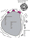
Dermatomyositis (DM) is a rare idiopathic inflammatory myopathy characterized by muscle weakness and cutaneous manifestations in adults and children. Calcinosis, a complication of DM, is the abnormal deposition of insoluble calcium salts in tissues, including skin, subcutaneous tissue, tendons, fascia, and muscle. Calcinosis is more commonly seen in juvenile DM (JDM), but also develops in adult DM. Although the mechanism of calcinosis remains unclear, several pathogenic hypotheses have been proposed, including intracellular accumulation of calcium secondary to an alteration of the cellular membrane by trauma and inflammation, local vascular ischemia, dysregulation of mechanisms controlling the deposition and solubility of calcium and phosphate, and mitochondrial damage of muscle cells. Identifying calcinosis biomarkers is important for early disease detection and risk assessment, and may lead to novel therapeutic targets for the prevention and treatment of DM-associated calcinosis. In this review, we summarize myositis autoantibodies associated with calcinosis in DM, histopathology and chemical composition of calcinosis, genetic and inflammatory markers that have been studied in adult DM and JDM-associated calcinosis, as well as potential novel biomarkers.
Keywords: Autoantibodies; Calcinosis; Dermatoyositis.
Published by Elsevier B.V.
Conflict of interest statement
Declaration of Competing Interest None.
Figures

References
-
- Muller S, Winkelmann R, Brunsting L. Calcinosis in dermatomyositis; observations on course of disease in children and adults. AMA Arch Derm. 1959;79(6):669–673. - PubMed
-
- Clemente G, Piotto DG, Barbosa C, et al. High frequency of calcinosis in juvenile dermatomyositis: a risk factor study. Rev Bras Reumatol. 2012;52(4):549–553. - PubMed
-
- Mathiesen P, Hegaard H, Herlin T, Zak M, Pedersen FK, Nielsen S. Long-term outcome in patients with juvenile dermatomyositis: a cross-sectional follow-up study. Scand J Rheumatol. 2012;41(1):50–58. - PubMed
Publication types
MeSH terms
Substances
Grants and funding
LinkOut - more resources
Full Text Sources

