. 2020 Mar 31;30(13):4490-4504.e4.
doi: 10.1016/j.celrep.2020.03.027.

Cortical Neural Stem Cell Lineage Progression Is Regulated by Extrinsic Signaling Molecule Sonic Hedgehog

Affiliations

Cortical Neural Stem Cell Lineage Progression Is Regulated by Extrinsic Signaling Molecule Sonic Hedgehog

Yue Zhang et al. Cell Rep. .

Abstract

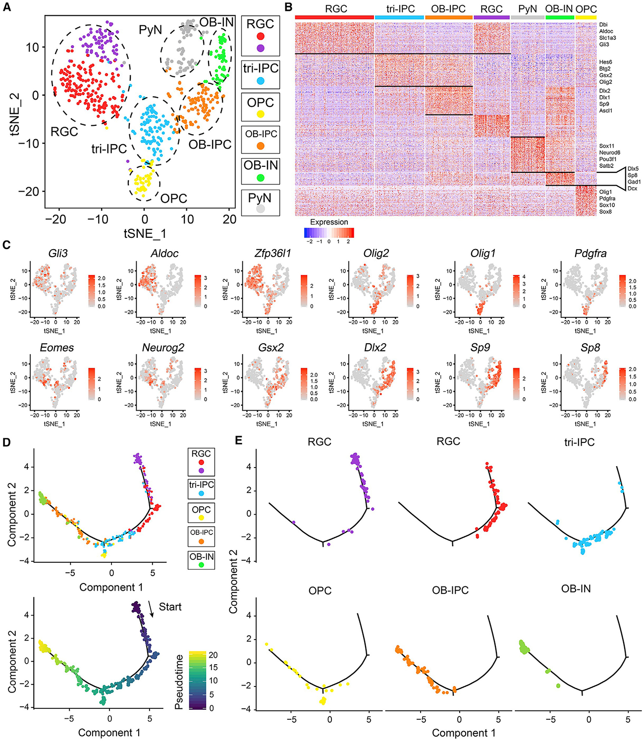
Neural stem cells (NSCs) in the prenatal neocortex progressively generate different subtypes of glutamatergic projection neurons. Following that, NSCs have a major switch in their progenitor properties and produce γ-aminobutyric acid (GABAergic) interneurons for the olfactory bulb (OB), cortical oligodendrocytes, and astrocytes. Herein, we provide evidence for the molecular mechanism that underlies this switch in the state of neocortical NSCs. We show that, at around E16.5, mouse neocortical NSCs start to generate GSX2-expressing (GSX2+) intermediate progenitor cells (IPCs). In vivo lineage-tracing study revealed that GSX2+ IPC population gives rise not only to OB interneurons but also to cortical oligodendrocytes and astrocytes, suggesting that they are a tri-potential population. We demonstrated that Sonic hedgehog signaling is both necessary and sufficient for the generation of GSX2+ IPCs by reducing GLI3R protein levels. Using single-cell RNA sequencing, we identify the transcriptional profile of GSX2+ IPCs and the process of the lineage switch of cortical NSCs.

Keywords: Gli3; Gsx2; Shh; cerebral cortex; neural stem cells; olfactory bulb interneurons; oligodendrocytes.


Conflict of interest statement

Declaration of Interests J.L.R. and A.R.K. are cofounders, stockholders, and on the scientific board of Neurona, a company studying the potential therapeutic use of interneuron transplantation. The other authors declare no competing interests.

Figures

Figure 1.
Figure 1.. Cortical NSCs Generate GSX2+ Tri-IPCs That Give Rise Not Only to OB Interneurons but Also to Cortical Oligodendrocytes and Astrocytes
(A) GSX2+ IPCs in the mouse cortical SVZ at E16.5, E18.5, and P5. Ctx, cortex; LV, lateral ventricle. (B) The strategy of the intersectional lineage analysis. (C) Plasmids pCAG-Cre were electroporated to the cortical VZ of Gsx2Flpo/+; IS mice at E14.5. GFP+ cells were observed in the SVZ and cortical plate at E18.5. CP, cortical plate; IZ, intermediate zone; MZ, marginal zone. (D) GFP+ and/or tdT+ interneurons in the OB at P21. (E) Quantification of the percentages of GFP+ and tdT+ cells among all the lineage-traced cells in the OB. (F–H) Cortical GFP+ oligodendrocytes (OLIG2+, arrows in G) and astrocytes (S100b+, arrows in H) with higher magnification images at P21. Note tdT+ PyNs located in cortical layers II–V (F). (I) Percentages of oligodendrocytes and astrocytes among all GFP+ cells in the cortex. Data in (E) and (I) were from three mice each. Scale bars, 50 mm in (A), (C), (D), and (F)–(H).
Figure 2.
Figure 2.. Cells of the OB Interneuron Lineage Are Not Generated in the Cortical SVZ of Smo cko Mice at P2
(A) In situ RNA hybridization showing expressions of Gli1, Gad1, Sp9, Tshz1, and Prokr2 in the caudal cortical SVZ (arrows) in the control and hGFAP-Cre; SmoF/F (Smo cko) mice. (B and C) GSX2 and SP8 (arrows) immunostainings of the rostral (B) and caudal (C) cortical sections from control and Smo cko mice at P2. Ctx, cortex; Str, striatum. (D) Numbers of GSX2+ and SP8+ cells in the cortical SVZ per section. Data are presented as means ± SEM; n = 3. ***p < 0.001, **p < 0.01; Student’s t test. Scale bars, 200 mm in (B) and (D).
Figure 3.
Figure 3.. Overexpression of ShhN in the Cortex by IUE Induces OB Interneuron and Oligodendrocyte Lineages in the Cortical SVZ
(A) Control pCAG-GFP plasmids (control-IUE) or pCAG-ShhN-ires-GFP plasmids (ShhN-IUE) were electroporated into the cortical VZ on E13.5. The E18.5 brains were analyzed. The distribution patterns of electroporated cells (GFP+) in the cortex are shown. Note that the mRNA levels of Gli1, Ptch1, Gad1, Tshz1, and Prokr2 were dramatically increased in the ShhN-IUE cortex. (B) The expressions of GSX2, ASCL1, DLX2, SP8, SP9, and OLIG2 were greatly increased in the ShhN-IUE cortex. (C) RNA-seq analysis revealed increased expression levels for SHH pathway target genes, OB interneuron lineage and oligodendrocyte lineage genes in the ShhN-IUE cortices at P0. Data are presented as means ± SEM; n = 3. ***p < 0.001, *p < 0.05; n.s., non-significant; Student’s t test in (C). Scale bars, 200 mm in (A) and (B).
Figure 4.
Figure 4.. SHH Regulates the Production of OB Interneurons and Oligodendrocytes in the Cortical SVZ Predominately by Reducing GLI3
(A–D) Immunostainings for GSX2, SP8, EOMES, and OLIG2 in wild-type (control) (A), Smo cko (B), Gli3 cko (C), and Smo Gli3 dcko (D) mice at P0. (E) Quantification for the numbers of GSX2+, SP8+, EOMES+, and OLIG2+ cells per 300 mm width in the cortical VZ-SVZ of control and mutant mice at P0. Data are presented as means ± SEM; n = 3 mice per genotype. *p < 0.05; unpaired Student’s t test in (E). Scale bars, 200 mm in (D).
Figure 5.
Figure 5.. scRNA-Seq Analysis of Cells in the E16.5 Wild-Type Cortices and in the ShhN-IUE Cortices
(A and B) Scatterplot of cells after principal-component analysis and t-SNE visualization, colored according to Seurat clustering and annotated by major cell types for all the cells in the wild-type sample (A) and the ShhN-IUE sample (B). (C and D) t-SNE of cells colored by mean expression of Gsx2 and Olig2 in wild-type (C) and ShhN-IUE (D) samples. (E) The eight Gsx2+ cells in the E16.5 wild-type sample consisted of four tri-IPCs and four OB-IPCs, based on the expressions of specific genes.
Figure 6.
Figure 6.. scRNA-Seq Analysis of the Progenitor Cells in the ShhN-IUE Sample
(A) Seurat clustering was performed on all the progenitor cells in the ShhN-IUE sample. Seven clusters were identified and annotated to six cell types based on gene expression features. (B) Heatmap showing marker gene expressions in the seven cell clusters. Each column represents expressions in one cell, and each row represents expressions of one gene. (C) The t-SNE plots of cells colored by mean expression of specific marker genes. (D) Monocle analysis of all the progenitors in the ShhN-IUE samples revealed differentiation trajectories and pseudo-timelines along the cell differentiation axis. Each point represents a cell, colored by cluster identity (top) or pseudo-timeline (bottom). (E) Seurat clusters shown along the predicted pseudo-timeline differentiation trajectory.
Figure 7.
Figure 7.. In Vivo Validation of Markers of Tri-IPCs and OB-IPCs
(A and B) The expression of GSX2, OLIG2, and DLX2 in the cortical VZ/SVZ of wild-type (A) and ShhN-IUE (B) mice at E17. Note that very few GSX2+ cells (green) were present in the cortical SVZ. Arrows indicate GSX2+OLIG2+DLX2 tri-IPCs, and arrowheads indicate GSX2+DLX2+ OB-IPCs. (C and D) The expression of SP9 and SP8 in the cortical VZ/SVZ of wild-type (C) and ShhN-IUE (D) mice at E17. (E and F) More OB interneuron lineage cells (E) and more tri-IPCs and OB-IPCs (F) were observed in the ShhN-IUE cortices than in the controls. (G) The sequential expression of GSX2/DLX2/SP9/SP8 is linked to lineage differentiation from tri-IPCs/OB-IPCs/OB neuroblasts, indicating the core transcriptional network for OB interneuron generation. Data are presented as means ± SEM; n = 3 mice for each condition. ***p < 0.001, **p < 0.01, *p < 0.05; Student’s t test in (E) and (F). Scale bars, 50 mm in (A)–(D) and (G).

References

    1. Anderson SA, Eisenstat DD, Shi L, and Rubenstein JL (1997). Interneuron migration from basal forebrain to neocortex: dependence on Dlx genes. Science 278, 474–476. - PubMed
    1. Balordi F, and Fishell G (2007). Hedgehog signaling in the subventricular zone is required for both the maintenance of stem cells and the migration of newborn neurons. J. Neurosci 27, 5936–5947. - PMC - PubMed
    1. Bayraktar OA, Fuentealba LC, Alvarez-Buylla A, and Rowitch DH (2014). Astrocyte development and heterogeneity. Cold Spring Harb. Perspect. Biol 7, a020362. - PMC - PubMed
    1. Blaess S, Stephen D, and Joyner AL (2008). Gli3 coordinates three-dimensional patterning and growth of the tectum and cerebellum by integrating Shh and Fgf8 signaling. Development 135, 2093–2103. - PMC - PubMed
    1. Blondel VD, Guillaume J-L, Lambiotte R, and Lefebvre E (2008). Fast unfolding of communities in large networks. J. Stat. Mech 10, P10008.

Publication types

MeSH terms