. 2020 May:390:107935.
doi: 10.1016/j.heares.2020.107935. Epub 2020 Mar 18.

Pericyte abnormalities precede strial capillary basement membrane thickening in Alport mice

Affiliations

Pericyte abnormalities precede strial capillary basement membrane thickening in Alport mice

Brianna Dufek et al. Hear Res. 2020 May.

Abstract

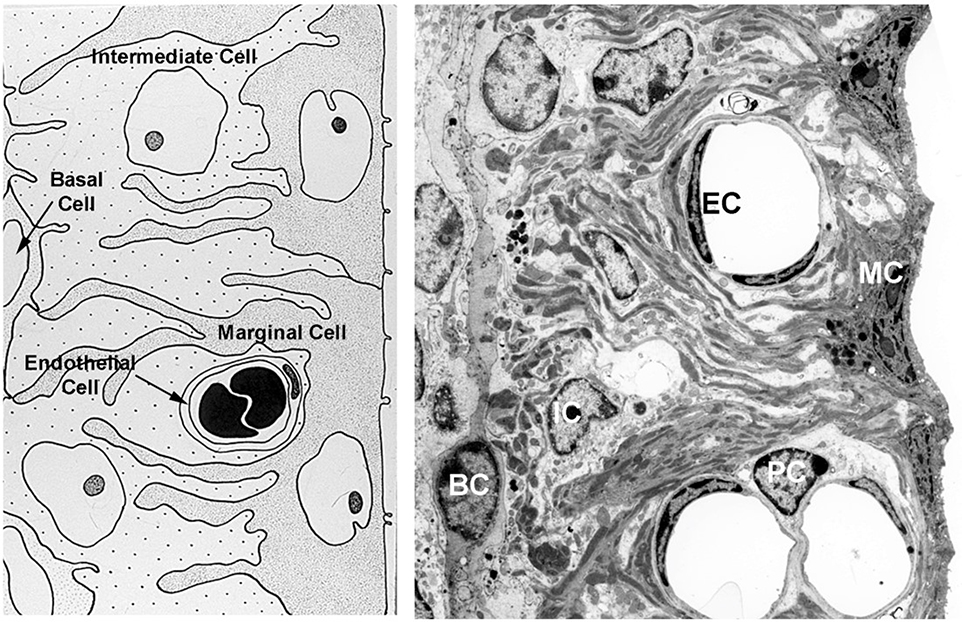
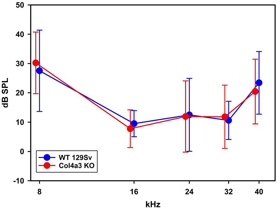
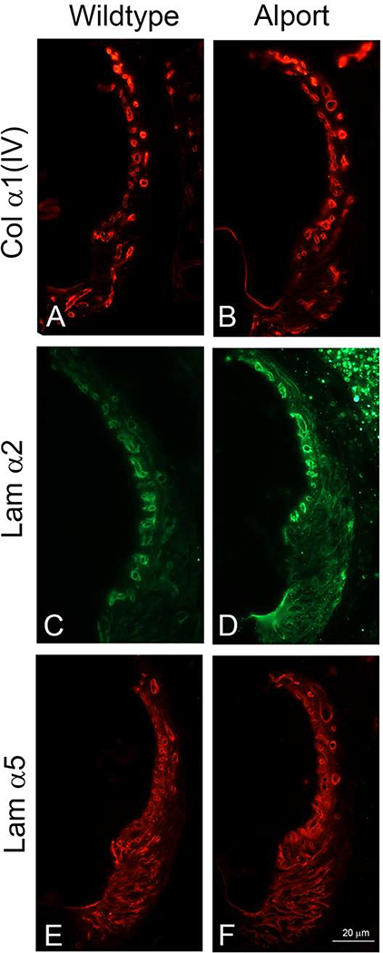
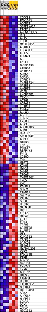
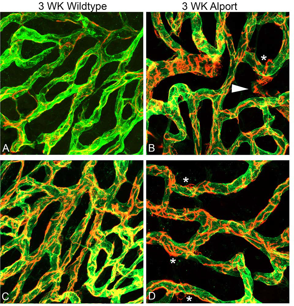
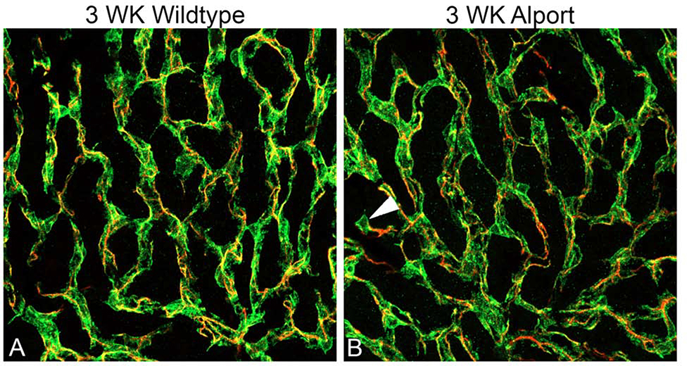
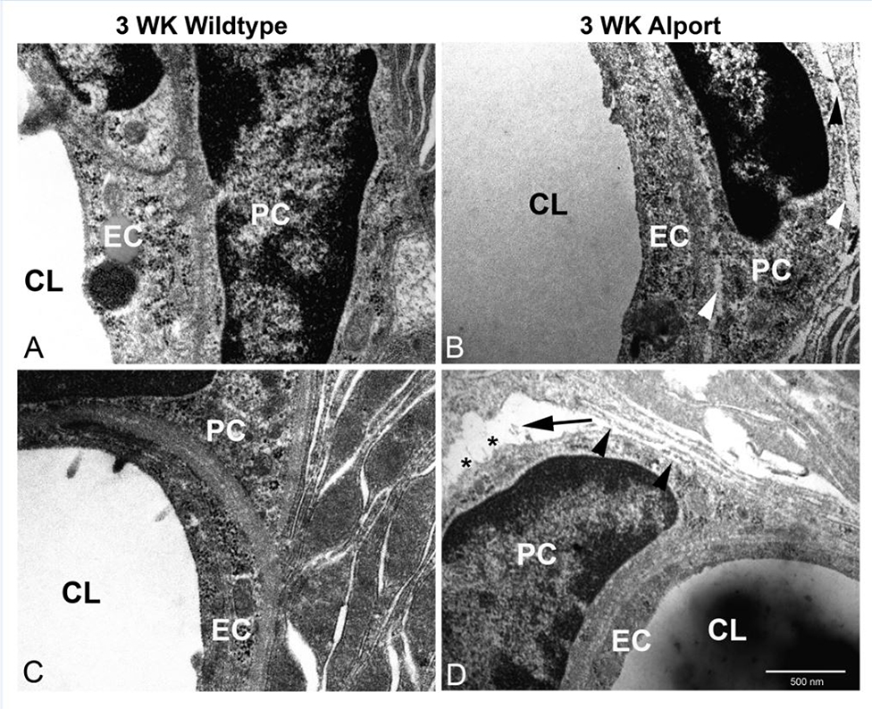
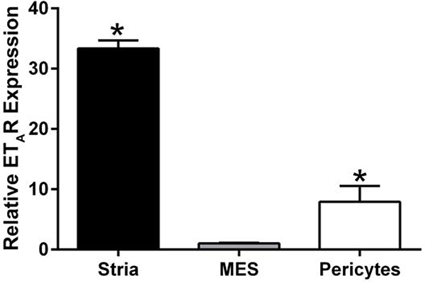
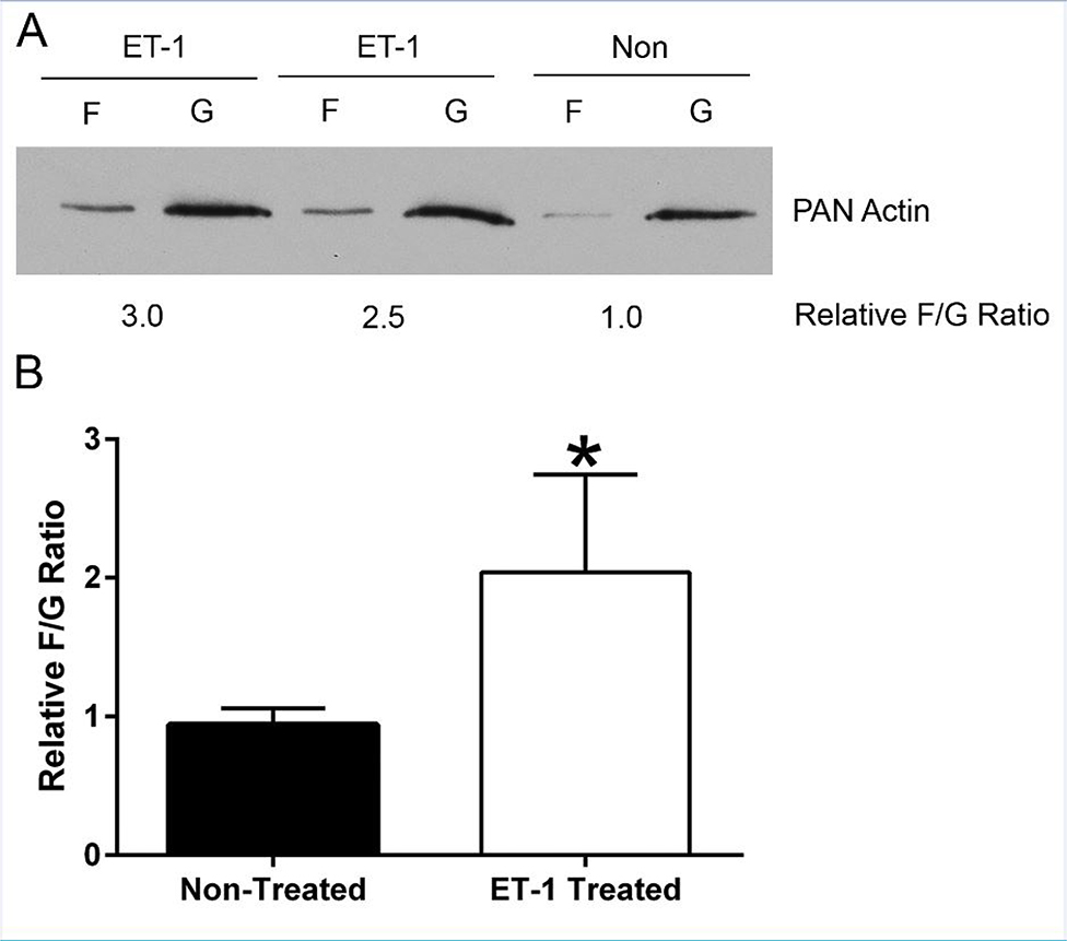
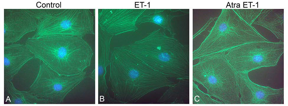
In 129 Sv autosomal Alport mice, the strial capillary basement membranes (SCBMs) progressively thicken between 5 and 9 weeks of age resulting in a hypoxic microenvironment with metabolic stress and induction of pro-inflammatory cytokines and chemokines. These events occur concomitant with a drop in endocochlear potential and a susceptibility to noise-induced hearing loss under conditions that do not permanently affect age/strain-matched littermates. Here we aimed to gain an understanding of events that occur before the onset of SCBM thickening. Alport stria has normal thickness and shows levels of extracellular matrix (ECM) molecules in the SCBMs commensurate with wild-type mice. Hearing thresholds in the 3-week Alport mice do not differ from those of wild-type mice. We performed RNAseq analysis using RNA from stria vascularis isolated from 3-week Alport mice and wild type littermates. Data was processed using Ingenuity Pathway Analysis software and further distilled using manual procedures. RNAseq analysis revealed significant dysregulation of genes involved in cell adhesion, cell migration, formation of protrusions, and both actin and tubulin cytoskeletal dynamics. Overall, the data suggested changes in the cellular architecture of the stria might be apparent. To test this notion, we performed dual immunofluorescence analysis on whole mounts of the stria vascularis from these same animals stained with anti-isolectin gs-ib4 (endothelial cell marker) and anti-desmin (pericyte marker) antibodies. The results showed evidence of pericyte detachment and migration as well as the formation of membrane ruffling on pericytes in z-stacked confocal images from Alport mice compared to wild type littermates. This was confirmed by TEM analysis. Earlier work from our lab showed that endothelin A receptor blockade prevents SCBM thickening and ECM accumulation in the SCBMs. Treating cultured pericytes with endothelin-1 induced actin cytoskeletal rearrangement, increasing the ratio of filamentous to globular actin. Collectively, these findings suggest that the change in type IV collagen composition in the Alport SCBMs results in cellular insult to the pericyte compartment, activating detachment and altered cytoskeletal dynamics. These events precede SCBM thickening and hearing loss in Alport mice, and thus constitute the earliest event so far recognized in Alport strial pathology.

Keywords: Alport syndrome; Pericyte; RNA-seq; Strial capillary basement membrane.


Conflict of interest statement

Declaration of competing interest The authors of this work have no conflicts of interest to disclose.

Figures

Figure 1.
Figure 1.
Cellular architecture of the stria vascularis. The stria consists of luminal marginal cells (MC) that form basolateral infolds with both intermediate cells (IC) and basal cells (BC). The capillaries are consisting of endothelial cells (IC) and pericytes (PC) which are both surrounded by SCBMs.
Figure 2.
Figure 2.
ABR thresholds indicate that 3-week-old Alport mice have normal hearing. ABR thresholds were determined for 3-week-old wild type and Alport mice. Variances for five individual measurements per cohort are shown. No significant differences were noted for any of the frequencies measured between cohorts (2-Way ANOVA, post-hoc Holm-Sidak).
Figure 3.
Figure 3.
Immunofluorescence analysis for ECM components in the SCBMs of wild type and Alport stria vascularis show no apparent differences. We previously showed that the SCBM thickening in Alport SCBMs was associated with accumulation of specific extracellular matrix molecules (Gratton et al., 2005; Meehan et al., 2016). This elevated expression is not observed in 3-week Alport mice compared to 3-week wild type mice. Data shown is representative of mid-modiolar sections from 3 animals per cohort. Lamα2 = laminin α2; Lama5 = laminin α5; Collagen IV = the α1 chain of collagen IV.
Figure 4.
Figure 4.
Heatmap of differentially expressed genes comparing wild type to Alport stria from 3 week old mice. The RNA-seq data was analyzed as described in the methods. The degree of red indicates higher relative expression and blue, lower relative expression across all individual samples analyzed.
Figure 5.
Figure 5.
Dual immunofluorescence immunostaining of whole mount cochlea for Alexa 488-labeled isolectin gs-beta4 and anti-desmin (red) from 3-week-old wild type (A and C) and Alport mice (B and D). Arrowhead: detached pericytes migrating into interstitium. Asterisks: pericytes ruffling. (63X magnification).
Figure 6.
Figure 6.
Dual immunofluorescence immunostaining of whole mount stria with anti-PDGFRb and anti-desmin from 3-week-old wild type (A) and Alport (B) mice. Pericyte somas are labeled with anti PDGFRb antibodies (green); pericyte filaments are labeled with anti-desmin antibodies (red) arrowhead indicates a migrating pericyte in the Alport sample. (40X magnification).
Figure 7.
Figure 7.
TEM analysis of pericyte detachment from the SCBMs in 3 week Alport stria. Strial capillaries from wild type mice show tight adherence between the pericytes and the epithelial/endothelial basement membranes (panels A and C). Alport strial capillaries occasionally show detachment Panels B and D, arrowheads) and less occasionally severe detachment (panel D arrow) with apparent membrane ruffling (panel D asterisks). These anomalies were never observed in the wild type strial capillaries. CL: capillary lumen; EC: endothelial cell; PC: pericyte.
Figure 8.
Figure 8.
Endothelin A receptors are expressed in both the stria vascularis and in cultured pericytes. RNA from the stria vascularis from wild type mice, cultured primary mesangial cells (positive control), and cultured primary pericytes were examined using real time RT-PCR. The experiment was performed in triplicate and analyzed by two-tailed student’s t-test. Asterisks denote significant differences (p<0.05) when compared to mesangial cell RNA (MES).
Figure 9.
Figure 9.
ET-1 effects on Actin Polymerization (F/G ratios) in Pericyte Cell Culture. Equal amounts of Pericytes cell lysates were subjected to differential centrifugation to separate filamentous actin (F-actin) from globular actin (G-actin) and Pan Actin immunoblots were performed from each sample (A). The corresponding bands were quantified and expressed as the F-actin/G-actin ratio. An increase of actin polymerization (F/G ratio) by ET-1 treatment was observed. This was quantified by densitometry for four independent experiments (B) using one-sample t test (hypothetical mean=1) * denotes a significant difference in F/G ratio for Et-1-treated versus non-treated cells.
Figure 10.
Figure 10.
ET-1 treatment of cultured pericytes results in an increase in actin filaments. Cells were treated vehicle (panel A), with ET-1 for 1 hour (panel B), or with a combination of ET-1 and the ETAR antagonist Atrasentan (panel C), and then fixed with acetone. Dual staining was performed using phalloidin (green) and anti-drebrin antibodies (red). Nuclei were stained with DAPI.

References

    1. Bertlich M, Ihler F, Weiss BG, Freytag S, Strupp M, Canis M 2017. Cochlear pericytes are capable of decreasing capillary diameter in vivo after tumor necrosis factor exposure. Otol. Neurotol 38, e545–e550. - PubMed
    1. Cosgrove D, Meehan DT, Grunkemeyer JA, Kornak JM, Sayers R, Hunter WT Samuelson, G.C. 1996. Collagen 4A3 knockout: A mouse model for autosomal Alport Syndrome. Genes and Development 10, 2981–2992. - PubMed
    1. Cosgrove DE, Samuelson G, Meehan DT, Miller C, McGee J, Walsh EJ & Siegel M 1998. Ultrastructural, physiological, and molecular defects in the inner ear of a gene-knockout mouse model for autosomal Alport syndrome. Hear. Res 121, 84–98. - PubMed
    1. Delimont D, Johnson BM, Meehan DT, Zallocchi M, Gratton MA,Phillips G, Cosgrove D 2014. Laminin 211-mediated FAK activation triggers Alport glomerular pathogenesis. PloS One 9(6):e99083. - PMC - PubMed
    1. Drees F, Pokutta S, Yamada S, Nelson WJ, Weis WI 2005. α-catenin is a molecular switch that binds E-cadherin-β-catenin and regulates actin filament assembly. Cell 123, 903–915. - PMC - PubMed

Publication types

MeSH terms