. 2020 Apr 14;117(15):8468-8475.
doi: 10.1073/pnas.1919960117. Epub 2020 Mar 31.

Identification of MLKL membrane translocation as a checkpoint in necroptotic cell death using Monobodies

Affiliations

Identification of MLKL membrane translocation as a checkpoint in necroptotic cell death using Monobodies

Emma J Petrie et al. Proc Natl Acad Sci U S A. .

Abstract

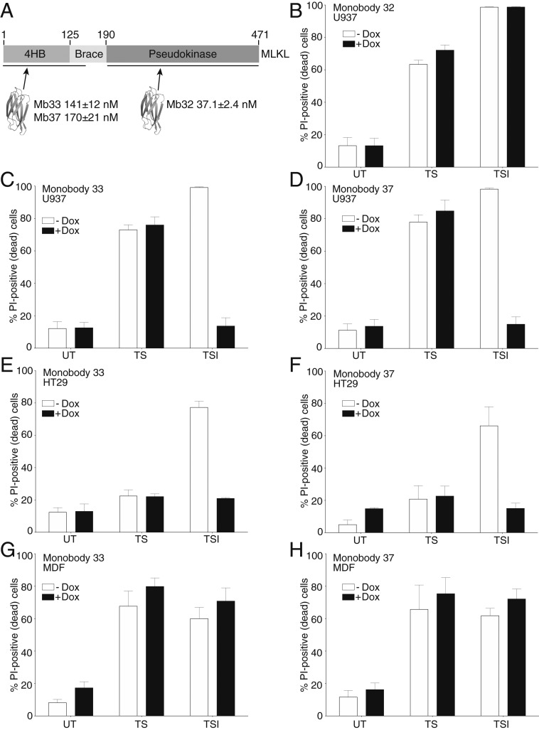
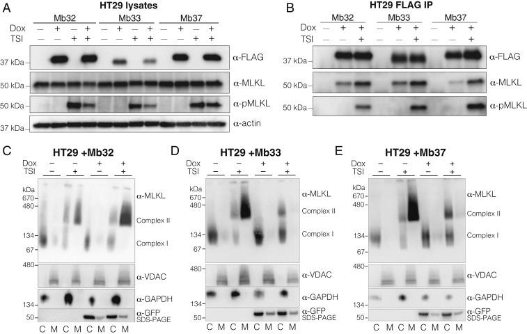
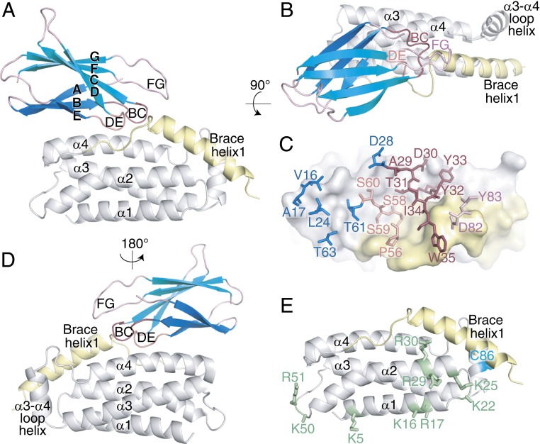
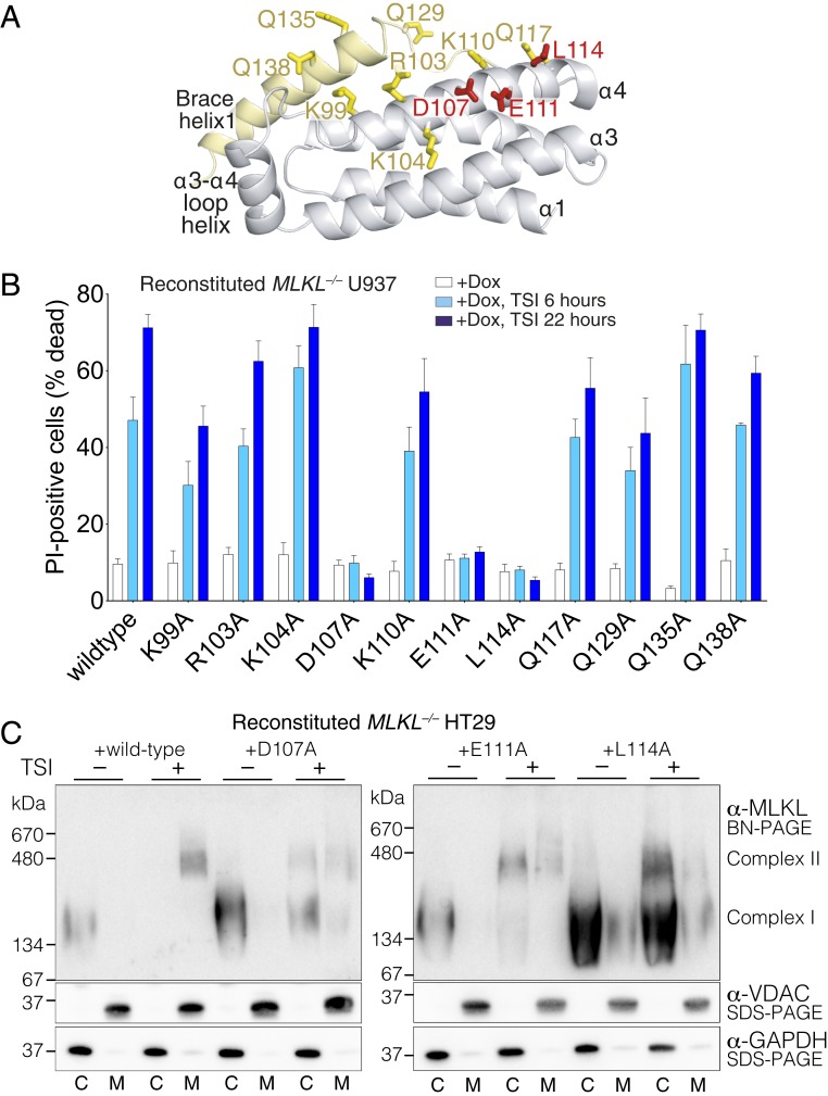
The necroptosis cell death pathway has been implicated in host defense and in the pathology of inflammatory diseases. While phosphorylation of the necroptotic effector pseudokinase Mixed Lineage Kinase Domain-Like (MLKL) by the upstream protein kinase RIPK3 is a hallmark of pathway activation, the precise checkpoints in necroptosis signaling are still unclear. Here we have developed monobodies, synthetic binding proteins, that bind the N-terminal four-helix bundle (4HB) "killer" domain and neighboring first brace helix of human MLKL with nanomolar affinity. When expressed as genetically encoded reagents in cells, these monobodies potently block necroptotic cell death. However, they did not prevent MLKL recruitment to the "necrosome" and phosphorylation by RIPK3, nor the assembly of MLKL into oligomers, but did block MLKL translocation to membranes where activated MLKL normally disrupts membranes to kill cells. An X-ray crystal structure revealed a monobody-binding site centered on the α4 helix of the MLKL 4HB domain, which mutational analyses showed was crucial for reconstitution of necroptosis signaling. These data implicate the α4 helix of its 4HB domain as a crucial site for recruitment of adaptor proteins that mediate membrane translocation, distinct from known phospholipid binding sites.

Keywords: RIPK3; cell death; programmed necrosis; protein engineering; protein interactions.


Conflict of interest statement

Competing interest statement: E.J.P., J.M.H., S.E.G., A.L.S., C.F., S.N.Y., P.E.C., and J.M.M. contribute to a project developing necroptosis inhibitors with Anaxis Pharma Pty Ltd. A.K. and S.K. are listed as inventors on issued and pending patents on the monobody technology filed by The University of Chicago (US Patent 9512199 B2 and related pending applications).

Figures

Fig. 1.
Fig. 1.
Monobodies targeting the human MLKL 4HB killer domain prevent necroptosis. (A) Scheme showing the domain architecture of MLKL with the domains targeted by monobodies developed in this work. The corresponding KD values are shown as mean ± SD of triplicate experiments. The impact of doxycycline-induced expression of Mb32, Mb33, and Mb37 in human U937 cells (B–D) and Mb33 and Mb37 in HT29 (E and F) and MDFs (G and H) was evaluated in untreated (UT), apoptotic (TS), or necroptotic (TSI) conditions. Cell death was measured by PI uptake and flow cytometry; death data represent mean ± SEM of three independent assays.
Fig. 2.
Fig. 2.
Mb33 and Mb37 inhibit necroptosis by blocking MLKL membrane translocation. Immunoblotting (A) and Mb immunoprecipitation (B) of lysates of HT29 cells with and without monobody expression. Mb33 and Mb37 did not prevent MLKL phosphorylation by RIPK3 following TSI stimulation. Expression of Mb32 (C), Mb33 (D), or Mb37 (E) in HT29 cells was induced by doxycycline (dox) treatment. Assembly of MLKL into higher-order species (“complex II”) and membrane translocation were assessed by blue-native PAGE ±7.5 h TSI treatment. Separation into cytoplasmic (“C”) and membrane (“M”) fraction was validated by blotting BN-PAGE for VDAC1 (membrane) and GAPDH (cytoplasmic). Expression of monobodies was verified by reducing SDS-PAGE blots for GFP. All images and blots are representative of two independent experiments.
Fig. 3.
Fig. 3.
The inhibitory monobody Mb33 blocks necroptosis by binding the human MLKL 4HB domain α4 helix and first brace helix. (A, B, and D) Transverse views of the Mb33:human MLKL 4HB domain-first brace helix cocrystal structure. Mb33 (β-sheets shown in teal) binds atop the 4HB domain (gray) α4 helix and the N-terminal portion of brace helix 1 (yellow). (C) Mb33 residues in FG (pink), BC (raspberry), and DE (salmon) loop and β-sheet (blue) located within 4.5 Å of the MLKL 4HB-brace helix (gray and yellow) are shown as sticks. (E) Residues previously implicated in phospholipid and inositol phosphate binding (green sticks) and the target of necrosulfonamide modification (C86; cyan sticks) are located on the opposing face to the monobody interaction interface (compare with A).
Fig. 4.
Fig. 4.
A functional site on the human MLKL 4HB domain α4 helix is crucial for MLKL to induce necroptotic death. (A) Cartoon of the 4HB domain-first brace helix structure rotated 30° about the x-axis relative to the depiction in Fig. 3D. MLKL residues proximal to Mb33 in the complex were selected for alanine substitution and are shown as sticks; essential residues for necroptotic signaling are shown as red sticks. (B) Effects of Ala substitutions of the indicated residues within full-length human MLKL on the capacity to reconstitute necroptotic signaling in MLKL/ human U937 cells. Wild-type or mutant MLKL expression was induced with doxycycline, and death was measured by PI uptake and flow cytometry in the absence of stimulus or the presence of TSI stimulation for 6 or 22 h. Exogenes were expressed in 2 to 3 independent MLKL−/− U937 clones (one clone for Q135A) and assayed independently to a combined n = 3 to 10 for each MLKL variant. Data are plotted as mean ± SEM. (C) Expression of wild-type, D107A, E111A, and L114A human MLKL in MLKL/ HT29 cells was induced by doxycycline (dox) treatment. Assembly of MLKL into higher-order species (“complex II”) and membrane translocation were assessed by blue-native PAGE ±7.5 h TSI treatment. Separation into cytoplasmic (“C”) and membrane (“M”) fractions was validated by blotting SDS-PAGE for VDAC1 (membrane) and GAPDH (cytoplasmic). All blots are representative of two independent experiments.

References

    1. Cho Y. S., et al. , Phosphorylation-driven assembly of the RIP1-RIP3 complex regulates programmed necrosis and virus-induced inflammation. Cell 137, 1112–1123 (2009). - PMC - PubMed
    1. Upton J. W., Kaiser W. J., Mocarski E. S., Virus inhibition of RIP3-dependent necrosis. Cell Host Microbe 7, 302–313 (2010). - PMC - PubMed
    1. Guo H., et al. , Herpes simplex virus suppresses necroptosis in human cells. Cell Host Microbe 17, 243–251 (2015). - PMC - PubMed
    1. Kitur K., et al. , Necroptosis promotes Staphylococcus aureus clearance by inhibiting excessive inflammatory signaling. Cell Rep. 16, 2219–2230 (2016). - PMC - PubMed
    1. Pearson J. S., et al. , EspL is a bacterial cysteine protease effector that cleaves RHIM proteins to block necroptosis and inflammation. Nat. Microbiol. 2, 16258 (2017). - PMC - PubMed

Publication types

Associated data

LinkOut - more resources