. 2020 Mar 31;13(625):eaay8248.
doi: 10.1126/scisignal.aay8248.

4-1BB costimulation promotes CAR T cell survival through noncanonical NF-κB signaling

Affiliations

4-1BB costimulation promotes CAR T cell survival through noncanonical NF-κB signaling

Benjamin I Philipson et al. Sci Signal. .

Abstract

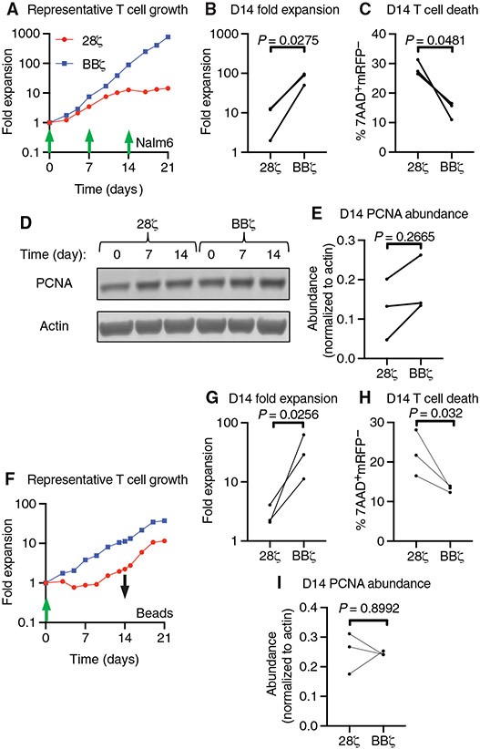
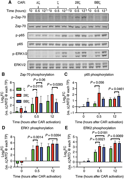
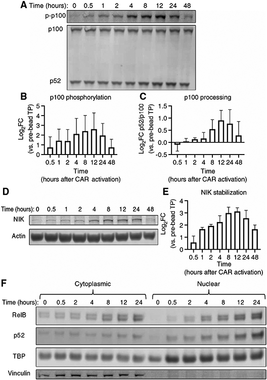
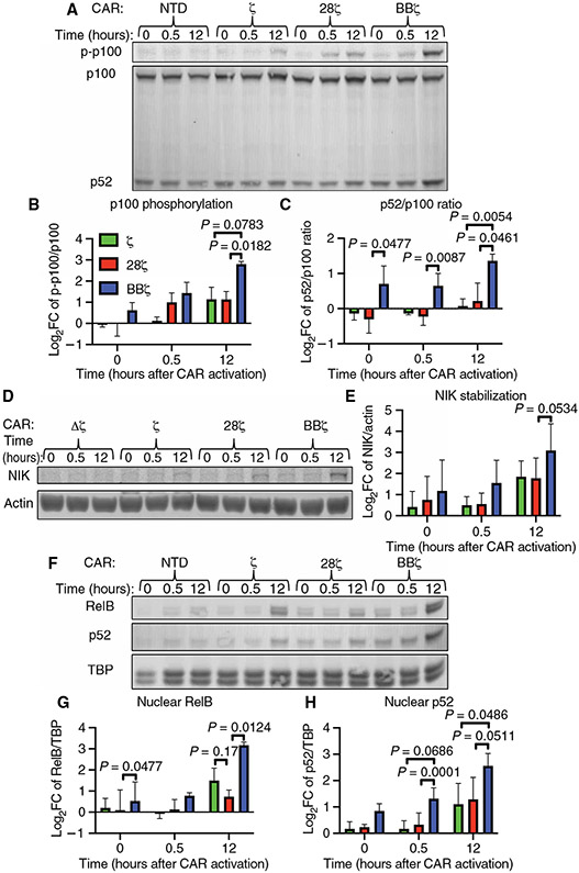
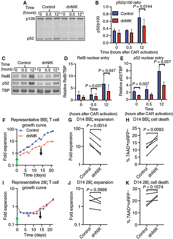
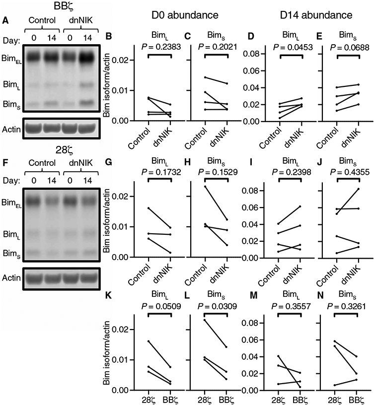
Clinical response to chimeric antigen receptor (CAR) T cell therapy is correlated with CAR T cell persistence, especially for CAR T cells that target CD19+ hematologic malignancies. 4-1BB-costimulated CAR (BBζ) T cells exhibit longer persistence after adoptive transfer than do CD28-costimulated CAR (28ζ) T cells. 4-1BB signaling improves T cell persistence even in the context of 28ζ CAR activation, which indicates distinct prosurvival signals mediated by the 4-1BB cytoplasmic domain. To specifically study signal transduction by CARs, we developed a cell-free, ligand-based activation and ex vivo culture system for CD19-specific CAR T cells. We observed greater ex vivo survival and subsequent expansion of BBζ CAR T cells when compared to 28ζ CAR T cells. We showed that only BBζ CARs activated noncanonical nuclear factor κB (ncNF-κB) signaling in T cells basally and that the anti-CD19 BBζ CAR further enhanced ncNF-κB signaling after ligand engagement. Reducing ncNF-κB signaling reduced the expansion and survival of anti-CD19 BBζ T cells and was associated with a substantial increase in the abundance of the most pro-apoptotic isoforms of Bim. Although our findings do not exclude the importance of other signaling differences between BBζ and 28ζ CARs, they demonstrate the necessary and nonredundant role of ncNF-κB signaling in promoting the survival of BBζ CAR T cells, which likely underlies the engraftment persistence observed with this CAR design.


Conflict of interest statement

Competing interests: C.H.J. reports receiving commercial research funding from Tmunity Therapeutics and has an ownership interest and intellectual property (IP) licensed to Novartis. M.C.M. reports receiving commercial research funding from Novartis and has an ownership interest and IP licensed to Cabaletta Bio. The other authors declare that they have no competing interests.

Figures

Fig. 1.
Fig. 1.. 4-1BB–costimulated CAR activation drives greater ex vivo T cell expansion and survival than does CD28-costimulated CAR activation.
(A) Representative ex vivo expansion of the indicated CAR T cells stimulated by irradiated Nalm6 target cells at the indicated times at a T cell to target cell ratio of 2:1. (B) Quantification of T cell expansion after 14 days of ex vivo culture from the experiments represented in (A). (C) Quantification of cell death by measurement of the percentage of 7AAD+mCherry cells in all events collected on day 14 of ex vivo culture from the experiments represented in (A). (D) Representative Western blotting analysis of PCNA abundance in the indicated CAR T cells at 0, 7, and 14 days of ex vivo culture with Nalm6 target cells. (E) Quantification of the relative amounts of PCNA in the indicated CAR T cells on day 14 of ex vivo culture with Nalm6 target cells. Data in (B), (C), and (E) are from three donors in two independent experiments and were analyzed by paired Student’s t test. (F) Representative ex vivo expansion of the indicated CAR T cells stimulated by anti-CD19 idiotype–coated target beads (at a T cell to bead ratio of 1:3) added at day 0 (green arrow) and removed at day 14 of culture (black arrow). (G) Quantification of T cell expansion after 14 days of ex vivo culture from the experiments represented in (F). (H) Quantification of cell death as assessed by measurement of the percentage of 7AAD+mCherry cells in all events collected on day 14 of ex vivo culture from the experiments represented in (F). Data in (G) and (H) are from three donors in three independent experiments and were analyzed by paired Student’s t test. (I) Quantification of the relative abundance of PCNA as assessed by Western blotting analysis of the indicated CAR T cells on days 0, 7, and 14 of ex vivo culture with target beads. Data are from donors in three independent experiments and were analyzed by paired Student’s t test.
Fig. 2.
Fig. 2.. Anti-CD19 idiotype target beads activate T cell signaling in anti-CD19 CAR T cells.
(A) Representative Western blotting of total and phosphorylated Zap-70, p65, and ERK1/2 proteins before and at 30 min and 12 hours after the addition of anti-CD19 beads to the indicated CAR T cells at a bead–to–T cell ratio of 5:1. The anti-CD19 CAR T cells used expressed CARs with no signaling domain (Δζ), the CD3ζ chain intracellular domain alone (ζ), the CD28 and CD3ζ chain intracellular domains (28ζ), or the 4-1BB and CD3ζ chain intracellular domains (BBζ). (B to E) Quantification of the relative abundances of phosphorylated Zap-70 (B), p65 (C), ERK1 (D), and ERK2 (E) normalized to their respective total proteins from the experiments represented in (A). P values were determined by two-way ANOVA with Tukey’s multiple comparisons test of data from three donors analyzed in three independent experiments. FC, fold change; TP, time point; NTD, non-transduced.
Fig. 3.
Fig. 3.. 4-1BB–costimulated anti-CD19 CAR activation stimulates CAR T cell ncNF-κB signaling.
(A) Representative Western blotting analysis of p100 phosphorylation and the processing of p100 to p52 at the indicated times before and after the addition of target beads to anti-CD19 BBζ CAR T cells. (B and C) Quantitative analyses of the relative amounts of phosphorylated p100 (B) (P = 0.0397 as determined by analyzing the data from two independent experiments using CAR T cells derived from three healthy donors by one-way ANOVA with the factor being time, as is the case for all of the P values in this figure) and the relative amounts of p52 generated by p100 processing (C) (p52/p100 ratio, P = 0.0244) from the experiments represented in (A). (D) Representative Western blotting analysis of NIK protein at the indicated times before and after target bead addition to anti-CD19 BBζ CAR T cells. Actin was used as a loading control. (E) Quantitative analysis of the relative amounts of NIK protein at the indicated times after the addition of beads to the cells in the experiments represented in (D) (P = 0.011). (F) Representative Western blotting analysis of the cytoplasmic and nuclear fractions from anti-CD19 BBζ CAR T cells before and at the indicated times after the addition of target beads. The blot is representative of two separate experiments using CAR T cells generated from two healthy donors (see fig. S6 for the other experiment).
Fig. 4.
Fig. 4.. 4-1BB, but not CD28, CAR costimulation drives basal and enhances CAR activation–induced ncNF-κB signaling.
(A) Representative Western blotting analysis of the phosphorylation of p100 and of its processing to p52 in the indicated CAR T cells at the indicated times before and after the addition of target beads at a bead–to–T cell ratio of 5:1. The anti-CD19 CARs used had no signaling domain (Δζ), the CD3ζ chain intracellular domain alone (ζ, green), the CD28 and CD3ζ chain intracellular domains (28ζ, red), or the 4-1BB and CD3ζ chain intracellular domains (BBζ, blue). (B and C) Quantitative analyses of the relative abundances of phosphorylated p100 (B) and of the ratio of p52 to p100 (C) from the experiments represented in (A). (D) Representative Western blotting analysis of NIK protein in the indicated CAR T cells at the indicated times before and after the addition of target beads as described for (A). Actin was used as a loading control. (E) Quantitative analysis of relative amounts of NIK protein in the indicated cells from the experiments represented in (D). (F) Representative Western blotting analysis of RelB, p52, and TBP (loading control) in nuclear fractions from the indicated anti-CD19 CAR T cells before and at the indicated times after the addition of target beads as described for (A). (G and H) Quantitative analyses of the relative amounts of nuclear RelB (G) and nuclear p52 (H) in the indicated cells from the experiments represented in (F). Data in (B), (C), (E), (G), and (H) are from three donors in three independent experiments. All log2(fold change relative to nonsignaling group) quantifications of band fluorescence intensities were normalized to those of the loading control. P values reported were determined by two-way ANOVA with Tukey’s multiple comparisons test.
Fig. 5.
Fig. 5.. Reducing 4-1BB costimulation–mediated ncNF-κB signaling diminishes BBζ CAR T cell ex vivo expansion and survival.
(A) Representative Western blotting analysis of p100 processing to p52 before, 30 min, and 12 hours after the addition of target beads to anti-CD19 BBζ CAR T cells expressing either mCherry (control) or mCherry and dnNIK. (B) Quantitative analysis of the ratio of p52 to p100 ratio from the experiments represented in (A). Data are from CAR T cells generated from three donors in three independent experiments. P value was determined by two-way ANOVA with Holm-Sidak’s multiple comparisons test. (C) Representative Western blotting analysis of RelB, p52, and TBP (loading control) in nuclear fractions of anti-CD19 BBζ CAR T cells treated as described for (A). (D and E) Quantitative analyses of the nuclear localization of RelB (D) and p52 (E) in cells from the experiments represented in (C) using TBP as the loading control. Data are from two independent experiments of CAR T cells generated from three donors. P values were determined by two-way ANOVA with Holm-Sidak’s multiple comparisons test. (F) Representative ex vivo expansion of control or dnNIK-expressing BBζ CAR T cells stimulated by anti-CD19 idiotype–coated target beads (at a T cell–to–bead ratio of 1:3) and added at day 0 (green arrow) and removed at day 14 of culture (black arrow). (G) Quantification of T cell expansion after 14 days of ex vivo culture from the experiments represented in (F). (H) Quantification of cell death by measurement of the percentage of 7AAD+mCherry cells in all events collected on day 14 of the ex vivo culture represented in (F). (I) Representative ex vivo expansion of control or dnNIK-expressing 28ζ CAR T cells stimulated by anti-CD19 idiotype–coated target beads at a T cell–to–bead ratio of 1:3 and added at day 0 (green arrow) and removed at day 14 of culture (black arrow). (J) Quantification of T cell expansion after 14 days of ex vivo culture from the experiments represented in (I). (K) Quantification of cell death by measurement of the percentage of 7AAD+mCherry cells in all events collected on day 14 of the ex vivo culture represented in (I). All P values were determined by paired Student’s t tests of data from four donors in four independent experiments.
Fig. 6.
Fig. 6.. 4-1BB costimulation–mediated ncNF-κB signaling opposes expression of the pro-apoptotic protein Bim.
(A) Representative Western blotting analysis of Bim isoforms in control and dnNIK-expressing BBζ CAR T cells before and 14 days after ex vivo expansion in response to the addition of beads. (B to E) Quantitation of the relative basal amounts of BimL (B) and BimS (C), as well as of the amounts of BimL (D) and BimS (E) 14 days after the addition of beads from the experiments represented in (A). Data in (B) to (E) are from four donors in four independent experiments. All P values were derived from paired Student’s t tests. (F) Representative Western blotting analysis of Bim isoforms in control or dnNIK-expressing 28ζ CAR T cells before and 14 days after ex vivo expansion in response to the addition of beads. (G and H) Quantitation of the relative basal amounts of BimL (G) and BimS (H) from the experiments represented in (F). Data are from three independent experiments with three donors. P values were derived from paired Student’s t tests. (I and J) Quantitation of the relative amounts of BimL (I) and BimS (J) 14 days after bead addition from the experiments represented in (F). Data in (I) and (J) are from four donors in four independent experiments. No pre-bead sample was taken for fourth donor shown in (I) and (J). P values were derived from paired Student’s t tests. (K and L) Comparison of the basal amounts of BimL (K) and BimS (L) between 28ζ and BBζ CAR T cells. (M and N) Comparison of the relative amounts of BimL (M) and BimS (N) between 28ζ and BBζ CAR T cells 14 days after expansion in response to beads. Data in (K) to (N) are from three donors in three independent experiments. All P values were derived from paired Student’s t tests.

References

    1. Feins S, Kong W, Williams EF, Milone MC, Fraietta JA, An introduction to chimeric antigen receptor (CAR) T-cell immunotherapy for human cancer. Am. J. Hematol 94, S3–S9 (2019). - PubMed
    1. Mueller KT, Waldron E, Grupp SA, Levine JE, Laetsch TW, Pulsipher MA, Boyer MW, August KJ, Hamilton J, Awasthi R, Stein AM, Sickert D, Chakraborty A, Levine BL, June CH, Tomassian L, Shah SS, Leung M, Taran T, Wood PA, Maude SL, Clinical pharmacology of tisagenlecleucel in B-cell acute lymphoblastic leukemia. Clin. Cancer Res 24, 6175–6184 (2018). - PMC - PubMed
    1. Orlando EJ, Han X, Tribouley C, Wood PA, Leary RJ, Riester M, Levine JE, Qayed M, Grupp SA, Boyer M, De Moerloose B, Nemecek ER, Bittencourt H, Hiramatsu H, Buechner J, Davies SM, Verneris MR, Nguyen K, Brogdon JL, Bitter H, Morrissey M, Pierog P, Pantano S, Engelman JA, Winckler W, Genetic mechanisms of target antigen loss in CAR19 therapy of acute lymphoblastic leukemia. Nat. Med 24, 1504–1506 (2018). - PubMed
    1. Mueller KT, Maude SL, Porter DL, Frey N, Wood P, Han X, Waldron E, Chakraborty A, Awasthi R, Levine BL, Melenhorst JJ, Grupp SA, June CH, Lacey SF, Cellular kinetics of CTL019 in relapsed/refractory B-cell acute lymphoblastic leukemia and chronic lymphocytic leukemia. Blood 130, 2317–2325 (2017). - PMC - PubMed
    1. Turtle CJ, Hanafi L-A, Berger C, Gooley TA, Cherian S, Hudecek M, Sommermeyer D, Melville K, Pender B, Budiarto TM, Robinson E, Steevens NN, Chaney C, Soma L, Chen X, Yeung C, Wood B, Li D, Cao J, Heimfeld S, Jensen MC, Riddell SR, Maloney DG, CD19 CAR-T cells of defined CD4+: CD8+composition in adult B cell ALL patients. J. Clin. Invest 126, 2123–2138 (2016). - PMC - PubMed

Publication types

MeSH terms

Substances