. 2020 Apr 1;12(7):5693-5715.
doi: 10.18632/aging.102958. Epub 2020 Apr 1.

Indian Hedgehog regulates senescence in bone marrow-derived mesenchymal stem cell through modulation of ROS/mTOR/4EBP1, p70S6K1/2 pathway

Affiliations

Indian Hedgehog regulates senescence in bone marrow-derived mesenchymal stem cell through modulation of ROS/mTOR/4EBP1, p70S6K1/2 pathway

Mahmoud Al-Azab et al. Aging (Albany NY). .

Abstract

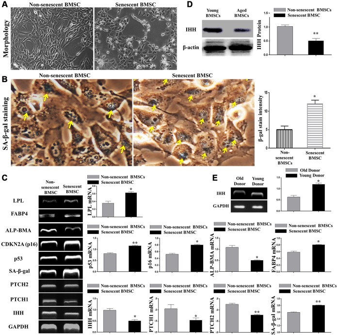
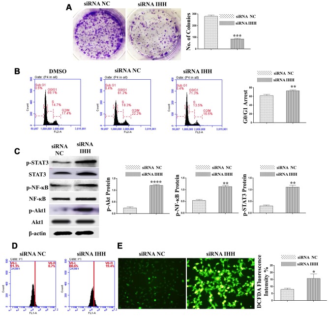
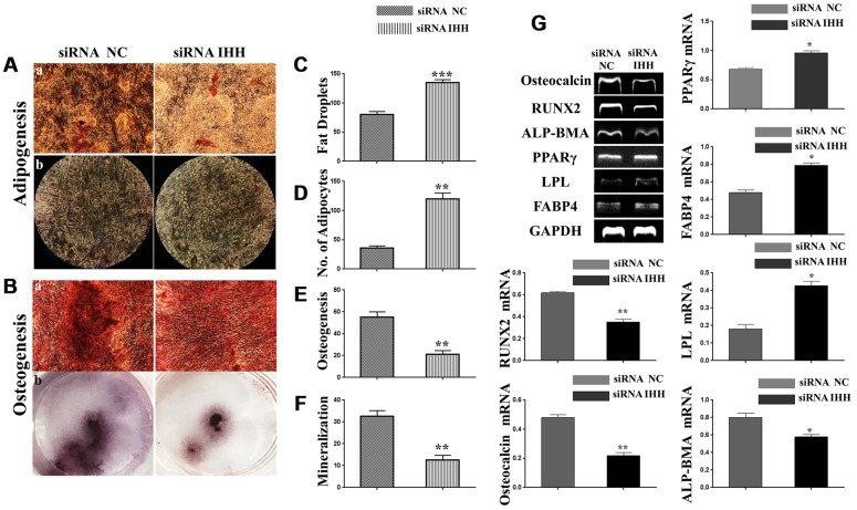
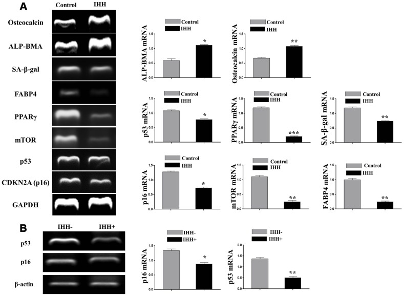
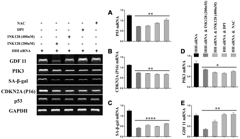
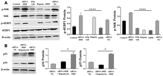
Premature senescence of bone marrow-derived mesenchymal stem cells (BMSC) remains a major concern for their application clinically. Hedgehog signaling has been reported to regulate aging-associated markers and MSC skewed differentiation. Indian Hedgehog (IHH) is a ligand of Hedgehog intracellular pathway considered as an inducer in chondrogenesis of human BMSC. However, the role of IHH in the aging of BMSC is still unclear. This study explored the role IHH in the senescence of BMSC obtained from human samples and senescent mice. Isolated BMSC were transfected with IHH siRNA or incubated with exogenous IHH protein and the mechanisms of aging and differentiation investigated. Moreover, the interactions between IHH, and mammalian target of rapamycin (mTOR) and reactive oxygen species (ROS) were evaluated using the corresponding inhibitors and antioxidants. BMSC transfected with IHH siRNA showed characteristics of senescence-associated features including increased senescence-associated β-galactosidase activity (SA-β-gal), induction of cell cycle inhibitors (p53/p16), development of senescence-associated secretory phenotype (SASP), activation of ROS and mTOR pathways as well as the promotion of skewed differentiation. Interestingly, BMSC treatment with IHH protein reversed the senescence markers and corrected biased differentiation. Moreover, IHH shortage-induced senescence signs were compromised after mTOR and ROS inhibition. Our findings presented anti-aging activity for IHH in BMSC through down-regulation of ROS/mTOR pathways. This discovery might contribute to increasing the therapeutic, immunomodulatory and regenerative potency of BMSC and introduce a novel remedy in the management of aging-related diseases.

Keywords: Indian hedgehog; aging; differentiation; mammalian target of rapamycin; mesenchymal stem cell.


Conflict of interest statement

CONFLICTS OF INTEREST: The authors declare no conflicts of interest.

Figures

Figure 1
Figure 1
IHH signaling pathway downregulated in aged BMSC. (A) Morphology of non-senescent and senescent BMSC. (B) Non-senescent and senescent BMSC stained by SA-β-gal stain. (C) BMSC (n = 4) were incubated with or without senescence-induction medium for 8 days. IHH, PTCH1/2, SA-β-gal, p53, p16, ALP-BMA, FABP4, and LPL genes expressions were measured by RT-PCR. GAPDH was used as a housekeeping gene. (D) Isolated BMSC (n = 4) were incubated with or without senescence-induction medium for 8 days. IHH protein expression was measured by Western Blot. β-actin was used as an internal control. (E) Isolated BMSC from young and old donors (n = 4) were analyzed for IHH gene expression by RT-PCR. GAPDH was used as a housekeeping gene. Results presented as mean ± SEM. *P <0.05, **P < 0.01.
Figure 2
Figure 2
IHH knockdown induced BMSC senescence. (A) BMSC transfected with NC siRNA and IHH siRNA stained by SA-β-gal stain. (B) BMSC (n = 5) were transfected with siRNA negative control or siRNA IHH for 24hours. TP53, CDKN2A, SA-β-gal, mTOR, PIK3, GDF11, COX-2, IDO1, IL-6, HGF, and TGFβ genes expressions were measured by RT-PCR. GAPDH was used as a housekeeping gene. (C) BMSC (n = 5) were transfected with siRNA negative control or siRNA IHH for 48hours. P53, P16, PI3K, and p-PI3K proteins expressions were measured by Western Blot. β-actin was used as an internal control. Results presented as mean ± SEM. *P <0.05, **P < 0.01, ***P < 0.001, ****P.
Figure 3
Figure 3
Silencing of IHH induced CFU inhibition, cell cycle arrest, and aging-related signaling pathways in BMSC. (A) BMSC (n = 5) were transfected for 24hours and then incubated in 10% FBS in MEM-ALPHA medium for 12 days. Colonies were visualized after staining by 0.02% crystal violet stain. (B) BMSC (n = 5) were transfected with siRNA negative control, siRNA IHH or DMSO for 24hours. Fixed cells stained by PI and RNase A, and then analyzed by flow cytometry for cell cycle distribution (C) BMSC (n = 5) were transfected with siRNA negative control or siRNA IHH for 48hours. Akt1, p-Akt1, NF-κB, p-NF-κB, STAT3, and p-STAT3 proteins expressions were measured by Western Blot. β-actin was used as an internal control. (D) BMSC (n = 5) were transfected with siRNA negative control or siRNA IHH for 24hours then stained by DCFHDA (5 μM). The fluorescent intensity of ROS was measured by flow cytometry and immunofluorescence microscopy (E). Results are shown as mean ± SEM. *P <0.05, **P < 0.01, ***P < 0.001, ****P < 0.0001.
Figure 4
Figure 4
Skewed differentiation was induced in IHH-depleted BMSC. (Aa, b), (C and D) BMSC (n = 5) were transfected with siRNA negative control or siRNA IHH for 24hours then incubated in adipogenesis differentiation medium for 21 days. Adipocytes were visualized under inverted light microscope and adipogenesis measured by staining fat droplets using Oil-Red-O stain. (Ba, b), (E, F) BMSC (n = 5) were transfected with siRNA negative control or siRNA IHH for 24hours then incubated in osteogenic differentiation medium for 21 days. Osteogenesis and Ca2 mineralization measured using Alizarine-Red-S stain. (G) RT-PCR for adipogenesis markers, PPARγ, LPL, and FABP4, and osteogenesis markers, ALP-BMA, RUNX2, and osteocalcin in BMSC with and without IHH siRNA. GAPDH was used as a housekeeping gene. Results have shown as mean ± SEM. *P <0.05, **P < 0.01, ***p <0.001.
Figure 5
Figure 5
IHH alleviated senescence and induced proper differentiation in BMSC. (A) Senescent BMSC (n=5) incubated with and without IHH for 24 hrs, then stained by SA-β-gal stain. (B) Non-senescent and senescent BMSC (n = 5) were incubated in osteogenic differentiation medium with and without IHH for 21 days. Osteogenesis measured using Alizarine-Red-S stain. (C) Non-senescent and senescent BMSC (n = 5) were incubated in adipogenesis differentiation medium with and without IHH for 21 days. Adipogenesis measured by staining fat droplets using Oil-Red-O stain. All data obtained are indicated as mean ± SEM. **P < 0.01, ***p <0.001, ****p <0.0001.
Figure 6
Figure 6
IHH reversed aging-related genes and promoted genes of proper differentiation. (A) BMSC (n = 5) were incubated with and without IHH for 24hours. Aging-related genes, TP53, CDKN2A, SA-β-gal, and mTOR, adipogenesis markers, PPARγ and FABP4, and osteogenesis markers, ALP-BMA, and osteocalcin genes expressions were measured by RT-PCR. GAPDH was used as a housekeeping gene. (B) BMSC from SAMP8 mice (n = 4) were incubated with and without IHH for 24hours. Aging-related genes, p16 and TP53 were measured by RT-PCR. β-actin was used as a housekeeping gene. All data obtained are indicated as mean ± SEM. *P <0.05, **P < 0.01, ***p <0.001.
Figure 7
Figure 7
IHH shortage-induced aging-associated genes suppressed by mTOR and ROS inhibition in BMSC. siRNA IHH-transfected BMSC (n=5) were incubated with or without INK128, DPI, or NAC for 24hours. TP53 (A), CDKN2A (B), SA-β-gal (C), PIK3 (D) and GDF11 (E) genes expressions were measured by RT-PCR. GAPDH was used as a housekeeping gene. All results were normally distributed and shown as mean ± SEM. *P <0.05, **P < 0.01, ***p <0.001, ****p <0.001.
Figure 8
Figure 8
mTOR and ROS inhibition reversed IHH depletion-induced senescence-related signaling pathways, cell cycle arrest, and inhibited CFU. siRNA IHH-transfected BMSC (n=5) were incubated with or without INK128, DPI, or NAC for 48hours. P53 (A), P16 (B), PI3K, p-PI3K (C), Akt1, p-Akt1 (D), NF-κB, p-NF-κB (E), STAT3, and p-STAT3 (F) proteins expressions were measured by Western Blot. β-actin was used as an internal control. (G) BMSC (n = 5) were treated with or without INK128, DPI, or NAC in presence of siRNA IHH for 24hours. Fixed cells stained by PI and RNase A, and then analyzed by flow cytometry for cell cycle distribution. (H) BMSC (n = 5) were treated with or without INK128, DPI, or NAC in presence of siRNA IHH for 24hours and then incubated in 10% FBS in MEM-ALPHA medium for 12 days. Colonies were visualized after staining with 0.02% crystal violet stain. All results were normally distributed and shown as mean ± SEM. *P <0.05, **p <0.01, ***p <0.001, ****p <0.001.
Figure 9
Figure 9
Biased differentiation induced by IHH shortage was corrected after mTOR and ROS inhibition in BMSC. (A) Morphology of siRNA IHH BMSC with and without INK128, DPI, or NAC. (Ba, b), (D, E) siRNA IHH-transfected BMSC (n=5) were incubated with or without INK128, DPI, or NAC for 24hours and then incubated in adipogenesis differentiation medium for 21 days. Adipocytes were visualized under inverted light microscope and adipogenesis measured by staining fat droplets using Oil-Red-O stain. (C and F) siRNA IHH-transfected BMSC (n=5) were incubated with or without INK128, DPI, or NAC for 24hours and then incubated in osteogenic differentiation medium for 21 days. Osteogenesis measured using Alizarine-Red-S stain. (G) RT-PCR for adipogenesis markers, PPARγ, LPL, and FABP4, and osteogenesis markers, ALP-BMA, RUNX2, and osteocalcin in IHH-depleted BMSC after incubation with or without INK128, DPI, or NAC for 24hours. GAPDH was used as a housekeeping gene. All results presented as mean ± SEM. *P <0.05, **P < 0.01, ***p <0.001, ****p <0.0001.
Figure 10
Figure 10
IHH inhibited phosphorylation of 4EBP1 and p70S6K1/2 in BMSC. (A) BMSC (n = 5) were transfected with siRNA negative control, siRNA IHH, rIHH, INK128, or rapamycin for 48hours. 4EBP1 and p70S6K1/2 total and phosphorylated proteins expressions were measured by Western Blot. β-actin was used as an internal control. (B) siRNA IHH-transfected BMSC (n=5) were incubated with or without rapamycin for 48hours. P16 and P53 proteins expressions were measured by Western Blot. β-actin was used as an internal control. All results were normally distributed and shown as mean ± SEM. *P <0.05, ****P<0.0001.

References

    1. Muller M. Cellular senescence: molecular mechanisms, in vivo significance, and redox considerations. Antioxid Redox Signal. 2009; 11:59–98. 10.1089/ars.2008.2104 - DOI - PubMed
    1. Duscher D, Rennert RC, Januszyk M, Anghel E, Maan ZN, Whittam AJ, Perez MG, Kosaraju R, Hu MS, Walmsley GG, Atashroo D, Khong S, Butte AJ, Gurtner GC. Aging disrupts cell subpopulation dynamics and diminishes the function of mesenchymal stem cells. Sci Rep. 2014; 4:7144. 10.1038/srep07144 - DOI - PMC - PubMed
    1. Bustos ML, Huleihel L, Kapetanaki MG, Lino-Cardenas CL, Mroz L, Ellis BM, McVerry BJ, Richards TJ, Kaminski N, Cerdenes N, Mora AL, Rojas M. Aging mesenchymal stem cells fail to protect because of impaired migration and antiinflammatory response. Am J Respir Crit Care Med. 2014; 189:787–98. 10.1164/rccm.201306-1043OC - DOI - PMC - PubMed
    1. Yukata K, Xie C, Li TF, Takahata M, Hoak D, Kondabolu S, Zhang X, Awad HA, Schwarz EM, Beck CA, Jonason JH, O’Keefe RJ. Aging periosteal progenitor cells have reduced regenerative responsiveness to bone injury and to the anabolic actions of PTH 1-34 treatment. Bone. 2014; 62:79–89. 10.1016/j.bone.2014.02.002 - DOI - PMC - PubMed
    1. Kaundal U, Bagai U, Rakha A. Immunomodulatory plasticity of mesenchymal stem cells: a potential key to successful solid organ transplantation. J Transl Med. 2018; 16:31. 10.1186/s12967-018-1403-0 - DOI - PMC - PubMed

Publication types

MeSH terms

Substances