

Invasive Stratified Mucin-producing Carcinoma (ISMC) of the Cervix: A Study on Morphologic Diversity
- PMID: 32235154
- PMCID: PMC7289664
- DOI: 10.1097/PAS.0000000000001480
Invasive Stratified Mucin-producing Carcinoma (ISMC) of the Cervix: A Study on Morphologic Diversity
Abstract
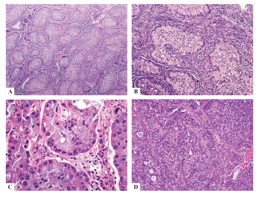
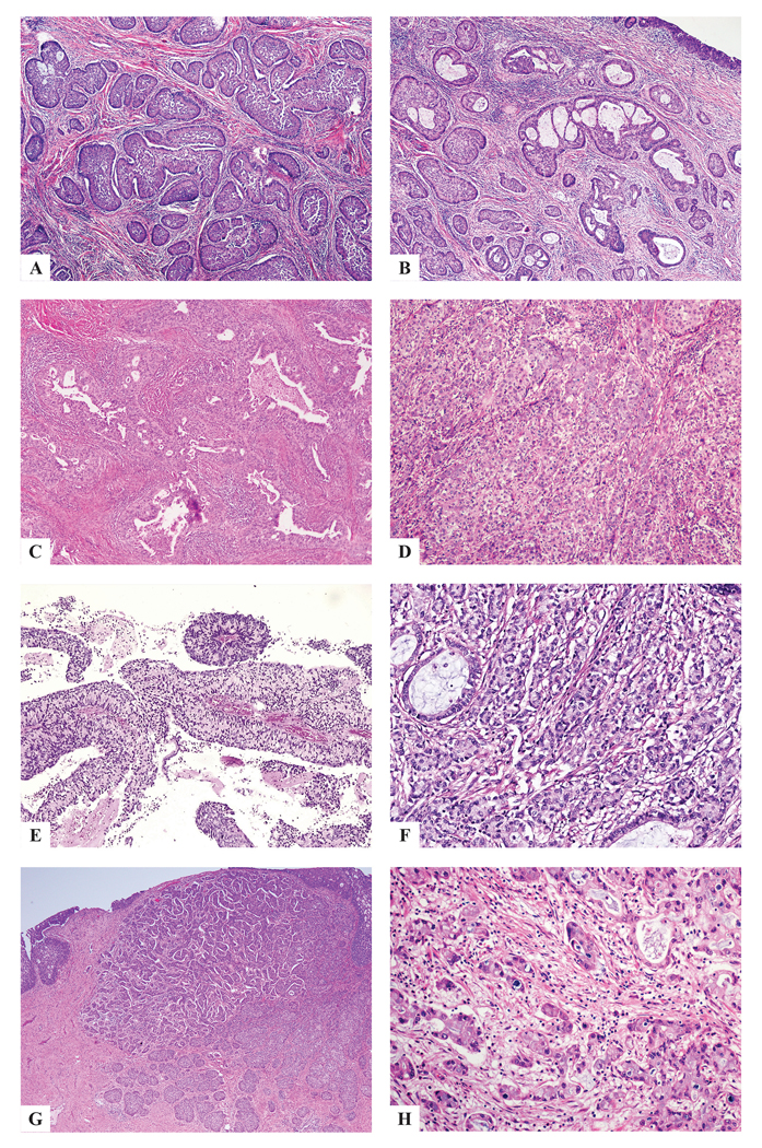
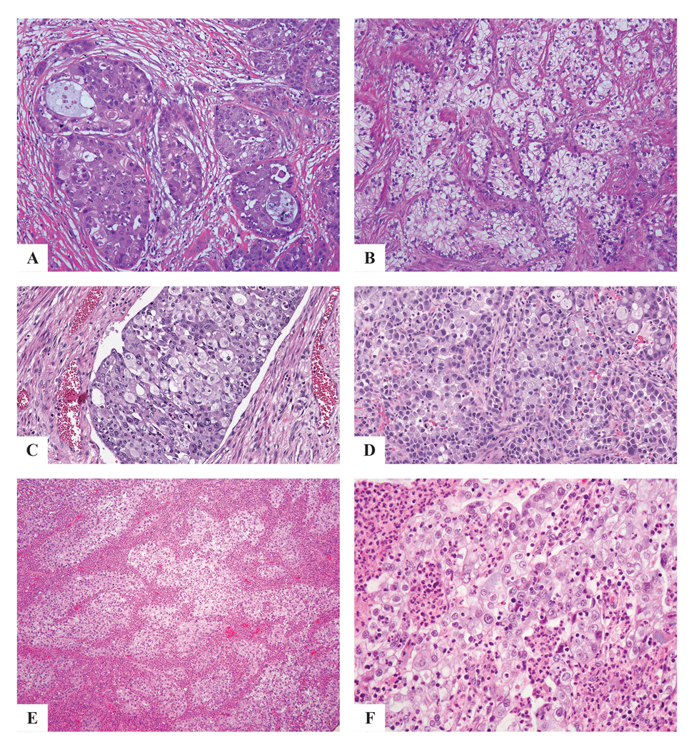
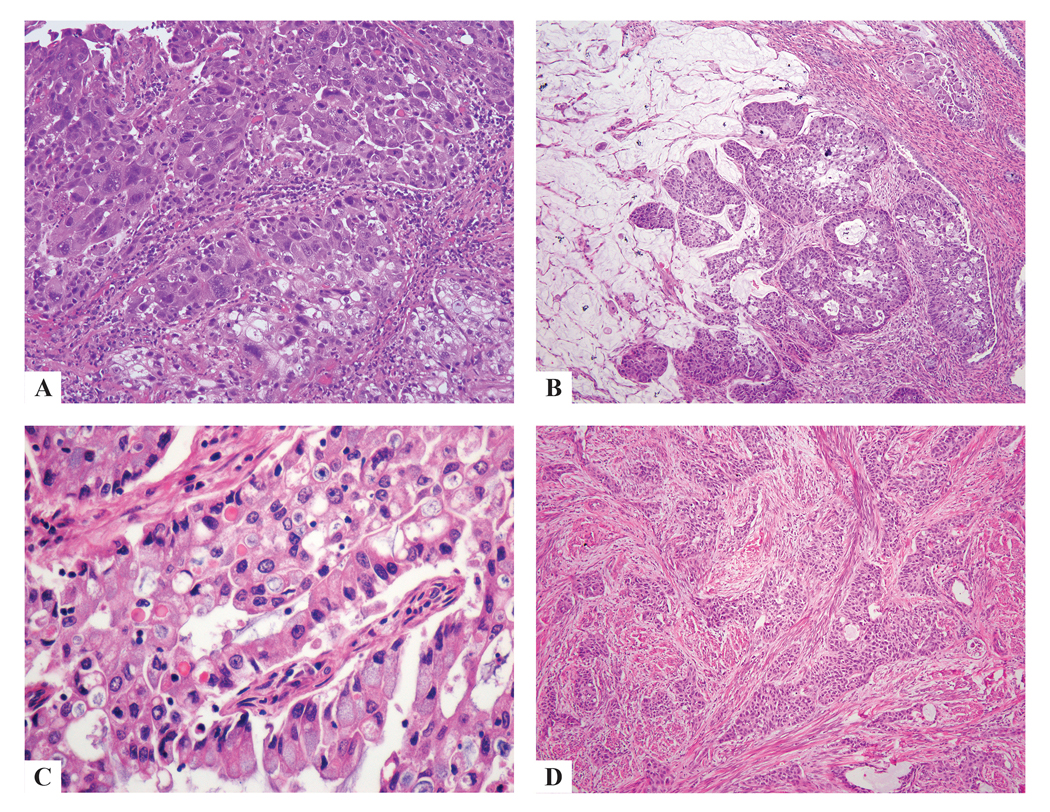
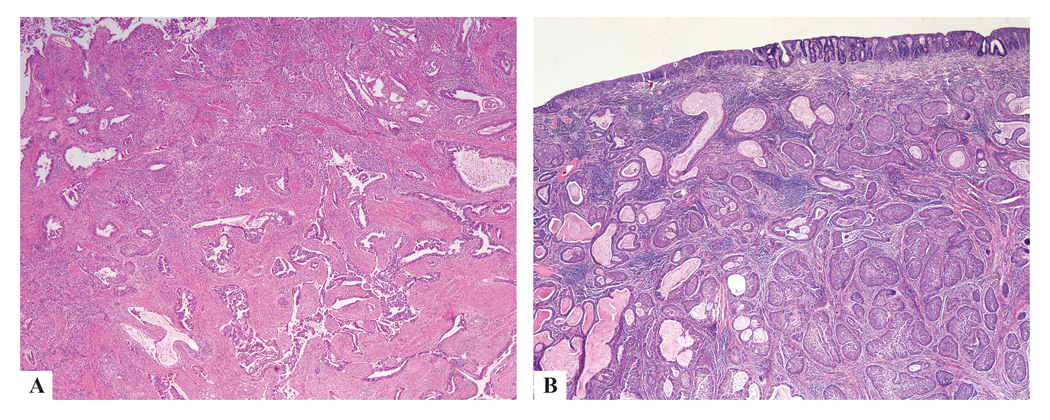
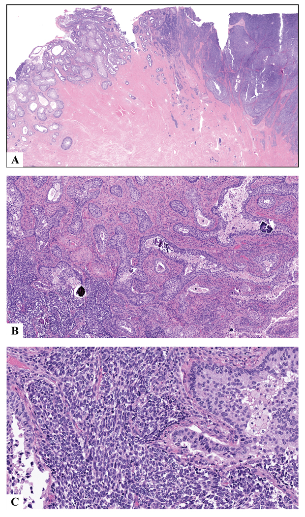
Invasive stratified mucin-producing carcinoma (ISMC) is a recently described tumor with similar morphology to the stratified mucin-producing intraepithelial lesion. Stratified mucin-producing intraepithelial lesion and ISMC likely arise from human papillomavirus (HPV)-infected reserve cells in the cervical transformation zone that retain their pluripotential ability to differentiate into various architectural and cytologic patterns. This is important, as small studies have suggested that ISMC may be a morphologic pattern associated with more aggressive behavior than usual HPV-associated adenocarcinoma. We sought to study the morphologic spectrum of this entity and its associations with other, more conventional patterns of HPV-associated carcinomas. Full slide sets from 52 cases of ISMC were reviewed by an international panel of gynecologic pathologists and classified according to the new International Endocervical Criteria and Classification system. Tumors were categorized as ISMC if they demonstrated stromal invasion by solid nests of neoplastic cells with at least focal areas of mucin stratified throughout the entire thickness, as opposed to conventional tall columnar cells with luminal gland formation. Tumors comprising pure ISMC, and those mixed with other morphologic patterns, were included in the analysis. Twenty-nine pure ISMCs (56%) and 23 ISMCs mixed with other components (44%) were identified. Other components included 13 cases of usual-type adenocarcinoma, 6 adenosquamous carcinoma, 3 mucinous-type adenocarcinoma, 1 high-grade neuroendocrine carcinoma. ISMC displayed architectural diversity (insular, lumen-forming, solid, papillary, trabecular, micropapillary, single cells) and variable cytologic appearance (eosinophilic cytoplasm, cytoplasmic clearing, histiocytoid features, glassy cell-like features, signet ring-like features, bizarre nuclei, squamoid differentiation). Awareness of the spectrum of morphologies in ISMC is important for accurate and reproducible diagnosis so that future studies to determine the clinical significance of ISMC can be conducted.
Conflict of interest statement
Figures
References
-
- Stoler M, Bergeron C, Colgan TJ, et al. Tumours of the Uterine Cervix In: Kurman RJ, Carcangiu ML, Herrington CS, et al., editors. WHO Classification of Tumours of Female Reproductive Organs, 4th Edition Lyon, France: IARC Press, 2014. p 184.
-
- Onishi J, Sato Y, Sawaguchi A, Yamashita A, Maekawa K, Sameshima H, Asada Y. Stratified mucin-producing intraepithelial lesion with invasive carcinoma: 12 cases with immunohistochemical and ultrastructural findings. Hum Pathol. 2016. September; 55:174–181. DOI: 10.1016/j.humpath.2016.05.007. - DOI - PubMed
