Review
. 2020 Mar 30;12(4):952.
doi: 10.3390/nu12040952.

The Role of Nutrition and Nutritional Supplements in Ocular Surface Diseases

Affiliations
Review

The Role of Nutrition and Nutritional Supplements in Ocular Surface Diseases

Marco Pellegrini et al. Nutrients. .

Abstract

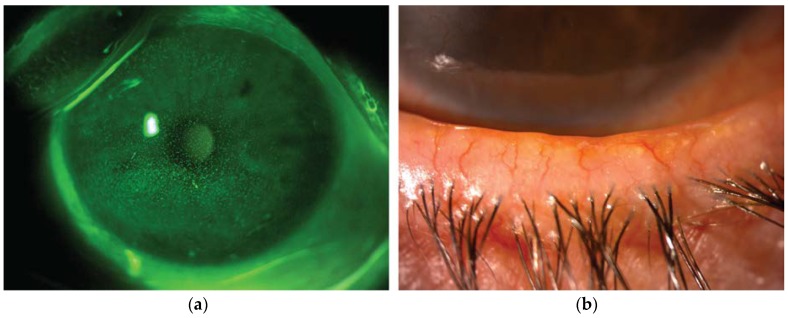
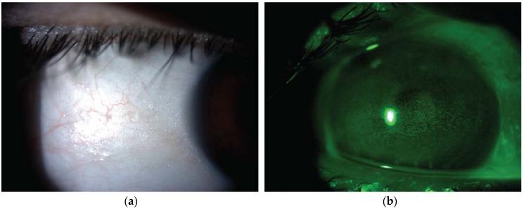
Dry eye disease (DED) is a multifactorial disease of the ocular surface system whose chore mechanisms are tear film instability, inflammation, tear hyperosmolarity and epithelial damage. In recent years, novel therapies specifically targeting inflammation and oxidative stress are being investigated and used in this field. Therefore, an increasing body of evidence supporting the possible role of different micronutrients and nutraceutical products for the treatment of ocular surface diseases is now available. In the present review, we analyzed in detail the effects on ocular surface of omega-3 fatty acids, vitamins A, B12, C, D, selenium, curcumin and flavonoids. Among these, the efficacy of omega-3 fatty acid supplementation in ameliorating DED signs and symptoms is supported by robust scientific evidence. Further long-term clinical trials are warranted to confirm the safety and efficacy of the supplementation of the other micronutrients and nutraceuticals.

Keywords: dry eye disease; nutraceuticals; nutritional supplements; ocular surface.


Conflict of interest statement

The authors declare no conflict of interest.

Figures

Figure 1
Figure 1
Representative image from a patient with dry eye disease owing to Meibomian gland dysfunction. (a) Slit lamp photograph of the cornea after instillation of 20 µL unpreserved 2% sodium fluorescein (corneal fluorescein staining). The epithelial damage is visible as multiple punctate epithelial erosions staining with fluorescein scattered over the corneal surface. (b) Slit lamp photograph of the lid margin showing typical signs of meibomian gland dysfunction, including hyperemia and irregularity of the lid margin, telangectasia, plugged gland orifices and anterior displacement of the mucocutaneous junction. Hyposecretion of meibomian lipids leads to tear film instability and increased evaporation rate.
Figure 2
Figure 2
Representative image from a patient with xerophthalmia due to vitamin A deficiency secondary to bariatric surgery. (a) Slit lamp photograph showing dry lusterless appearance of the keratinized bulbar conjunctiva (Bitot spot). (b) Corneal fluorescein staining showing multiple punctate epithelial erosions within the palpebral fissure.

References

    1. Gipson I.K. The ocular surface: The challenge to enable and protect vision: The Friedenwald lecture. Investig. Opthalmol. Vis. Sci. 2007;48:4391–4398. doi: 10.1167/iovs.07-0770. - DOI - PMC - PubMed
    1. Aragona P., Rolando M. Towards a dynamic customised therapy for ocular surface dysfunctions. Br. J. Ophthalmol. 2013;97:955–960. doi: 10.1136/bjophthalmol-2012-302568. - DOI - PubMed
    1. Stapleton F., Alves M., Bunya V.Y., Jalbert I., Lekhanont K., Malet F., Na K.-S., Schaumberg D., Uchino M., Vehof J., et al. TFOS DEWS II Epidemiology Report. Ocul. Surf. 2017;15:334–365. doi: 10.1016/j.jtos.2017.05.003. - DOI - PubMed
    1. Miljanović B., Dana R., Sullivan D.A., Schaumberg D.A. Impact of Dry Eye Syndrome on Vision-Related Quality of Life. Am. J. Ophthalmol. 2007;143:409–415. doi: 10.1016/j.ajo.2006.11.060. - DOI - PMC - PubMed
    1. Wolffsohn J.S., Arita R., Chalmers R., Djalilian A., Dogru M., Dumbleton K., Gupta P.K., Karpecki P., Lazreg S., Pult H., et al. TFOS DEWS II Diagnostic Methodology report. Ocul. Surf. 2017;15:539–574. doi: 10.1016/j.jtos.2017.05.001. - DOI - PubMed

MeSH terms