Review
. 2020 Mar 30;9(4):831.
doi: 10.3390/cells9040831.

Mitophagy in the Pathogenesis of Liver Diseases

Affiliations
Review

Mitophagy in the Pathogenesis of Liver Diseases

Po-Yuan Ke. Cells. .

Abstract

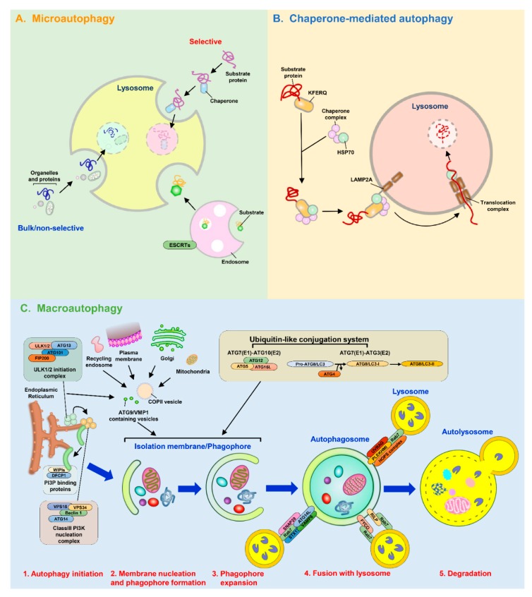
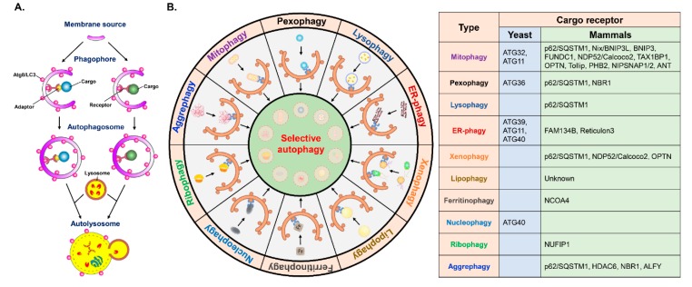
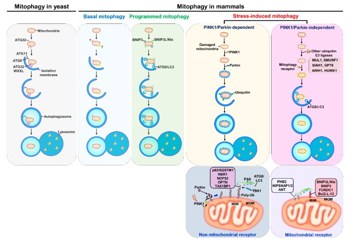
Autophagy is a catabolic process involving vacuolar sequestration of intracellular components and their targeting to lysosomes for degradation, thus supporting nutrient recycling and energy regeneration. Accumulating evidence indicates that in addition to being a bulk, nonselective degradation mechanism, autophagy may selectively eliminate damaged mitochondria to promote mitochondrial turnover, a process termed "mitophagy". Mitophagy sequesters dysfunctional mitochondria via ubiquitination and cargo receptor recognition and has emerged as an important event in the regulation of liver physiology. Recent studies have shown that mitophagy may participate in the pathogenesis of various liver diseases, such as liver injury, liver steatosis/fatty liver disease, hepatocellular carcinoma, viral hepatitis, and hepatic fibrosis. This review summarizes the current knowledge on the molecular regulations and functions of mitophagy in liver physiology and the roles of mitophagy in the development of liver-related diseases. Furthermore, the therapeutic implications of targeting hepatic mitophagy to design a new strategy to cure liver diseases are discussed.

Keywords: autophagy; fibrosis; hepatitis; hepatocellular carcinoma; liver disease; liver injury; mitophagy; steatosis.


Conflict of interest statement

Funding: This research was funded by the National Health Research Institute (NHRI-EX103-10322SC, NHRI-EX104-10322SC, NHRI-EX105-10322SC, and NHRI-EX106-10322SC), Miaoli; the Ministry of Science and Technology (MOST 102-2320-B-182-037-MY3, MOST 105-2628-B-182-001-MY3, and MOST108-2320-B-182-011), Taipei; and Chang Gung Memorial Hospital (CMRPD1C0211, CMRPD1D0021, CMRPD1D0022, CMRPD1D0023, CMRPD1G0281, CRRPD1F0031, CRRPD1F0032, CRRPD1F0033 and CMRPD1H0681), Taoyuan, Taiwan.

Figures

Figure 1
Figure 1
Overview of the molecular mechanisms regulating autophagy. Autophagy is classified into three major types: microautophagy, chaperone-mediated autophagy (CMA), and macroautophagy. (A) Microautophagy is a dynamic lysosomal membrane process that directly enwraps and deliver the intracellular portions to the lumen of lysosomes for degradation. (B) CMA is a lysosomal degradation process that involves the recognition of proteins containing the KFERQ-like motif by HSC70 and transport into the lysosomal lumen via the interaction with LAMP2A. (C) Macroautophagy (referred to here as autophagy) is a degradation pathway that promotes membrane rearrangement to generate vacuoles to engulf the cytosolic components targeted for degradation within lysosome. Two distinct metabolic sensors, AMPK and the mTOR complex, control the initiation of autophagy. Nutrient starvation in cells triggers the suppression of mTOR by AMPK1and the translocation of the ULK1/2 complex (ULK1/2, ATG13, RB1-inducible coiled-coil 1 (RB1CC1, also known as FIP200) and ATG101) to the autophagy initiation site. Subsequently, the ULK1/2 complex leads to the recruitment and activation of the class III phosphatidylinositol-3-OH kinase (class III-PI3K complex, including Vps34/PI3KC3, Vps15, Beclin 1, and ATG14) to synthesize PtdIn(3)P. The generated PtdIn(3)P then recruits DFCP1 and WIPI family proteins to the ER-associated membrane compartment to induce the formation of the isolation membrane (IM)/phagophore. In addition to the ER, other organelles, such as the plasma membrane, mitochondria, Golgi apparatus, and recycling endosome, also supply membrane resources required for the membrane nucleation and the formation of phagophore in the initial step of autophagy. ATG9, VMP1, and coated protein complex-associated vesicles are involved in the trafficking of membrane resources for the formation of the phagophore. The expansion and elongation of the phagophore to form enclosed autophagosomes rely on two ubiquitin-like (UBL) conjugation systems. First, ATG12 is conjugated to ATG5 via ATG7 (ubiquitin activating enzyme 1, E1) and ATG10 (ubiquitin conjugation enzyme 2, E2), yielding an ATG5-ATG12 conjugate that binds to ATG16L to form an ATG5-ATG12-ATG16L complex. Second, ATG8/LC3 family proteins are post-translationally processed by a cysteine protease ATG4 to form ATG8/LC3-I. Then, ATG7 (E1) and ATG3 (E2) enzyme cascades mediate the covalent linkage of ATG8/LC3-I to PE to form the lipidated form of LC3 (ATG8/LC3-PE, also known as ATG8/LC3-II). The enclosed autophagosomes fuse with lysosomes to form mature autolysosomes, which eliminate the engulfed materials. The interactions of RAB) with FYCO1 and RILP regulate the fusion of autophagosomes with lysosomes. In addition, several protein-protein interactions and the assembly of protein complexes, including the interaction between UVRAG and RAB7, the association of PLEKHM1 with the HOPS complex, and the formation of a protein complex containing ATG14L, STX17, SNAP29, and VAMP8, also regulate autophagosome-lysosome fusion.
Figure 2
Figure 2
Regulation of selective autophagy by cargo receptors. (A) Selective autophagy is an elimination process that involves the targeting of cargoes to the autophagy machinery by the specific receptor proteins that contain an ATG8/LC3-interacting regions (LIRs) for the interaction with ATG8/LC3 located on the membrane of the IM/phagophore. Another type of selective autophagy is mediated by the interactions between adaptor proteins with cargo receptors and ATG8/LC3. The IM/phagophore encloses mature autophagosomes that fuse with lysosomes to form autolysosomes, in which the engulfed cargoes are degraded. Ubiquitination of cargo and/or the binding to additional adaptor proteins mediate the recognition of cargoes and cargo receptors. (B) Selective autophagy participates in the degradation of damaged organelles and aggregated proteins. Several mammalian and yeast cargo receptors have been identified to eliminate the dysfunctional mitochondria, damaged lysosomes, injured peroxisomes, stressed ER and infecting pathogens by selective autophagy (mitophagy, lysophagy, pexophagy, ER-phagy, and xenophagy, respectively). Moreover, other forms of cargoes, including LDs (lipophagy), ferritin (ferritinophagy), nucleus (nucleophagy), ribosome (ribophagy), protein aggregate (aggrephagy), are degraded by selective autophagy through a process mediated by identified and unknown cargo receptors. The yeast and mammalian cargo receptors responsible for each type of selective autophagy are listed in the table.
Figure 3
Figure 3
Different types of mitophagy in yeast and mammals. In yeast cells, the mitochondrial outer membrane-associated protein ATG32 represents a receptor involved in mitophagy that interacts with ATG8 located on IM/phagophore through the WXXL-like motif. Another adaptor protein, ATG11, may mediate the interaction between ATG32 and ATG8 to facilitate mitophagy initiation. At least three types of mitophagy have been in mammals, including basal mitophagy, programmed mitophagy, and stress-induced mitophagy. Basal mitophagy has been shown to be activated in the tissues with large metabolic demands, but the specific receptor for basal mitophagy remains unclear. The cargo receptors of programmed mitophagy, including BNIP3 and BNIP3L/Nix, function in mitochondrial turnover for the maturation of erythrocytes and cardiomyocytes, and elimination of the parental mitochondrial genome during fertilization. Stress-induced mitophagy is classified into PINK1/Parkin-dependent mitophagy and PINK1/Parkin-independent mitophagy. For the activation of PINK1/Parkin-dependent mitophagy, mitochondrial depolarization triggers the suppression PINK1 degradation and leads to PINK1 accumulation on the MOM of damaged mitochondria. In turn, PINK1 phosphorylates ubiquitin and Parkin at serine residue 65 and then promotes the mitochondrial translocation of Parkin and mitochondrial ubiquitination by Parkin. On the other hand, several ubiquitin E3 ligases, including MUL1, SMURF1, SIAH1, GP78, ARIH1, and HUWE1, may promote the ubiquitination of mitochondrial proteins to activate PINK1/Parkin-independent mitophagy. Several non-mitochondrial cargo receptors, including p62/SQSTM1, Calcoco2 (also known as NDP52), OPTN, NBR1, and TAX1BP1, are responsible for PINK1/Parkin-dependent mitophagy. The phosphorylation of cargo receptors by tank-binding kinase 1, such as OPTN and p62/SQSTM1, facilitate the recognition process of mitophagy. Several MOM- and MIM-associated proteins, such as BNIP3, BNIP3L/Nix, FUNDC1, BCL2-L3, PHB2, ANT complex, and NIPSNAP1 and 2 have been identified as cargo receptors for mitophagy that directly bind to ATG8/LC3 located on the IM/phagophore.

References

    1. Levine B., Packer M., Codogno P. Development of autophagy inducers in clinical medicine. J. Clin. Investig. 2015;125:14–24. doi: 10.1172/JCI73938. - DOI - PMC - PubMed
    1. Choi A.M., Ryter S.W., Levine B. Autophagy in human health and disease. N. Engl. J. Med. 2013;368:651–662. doi: 10.1056/NEJMra1205406. - DOI - PubMed
    1. Gatica D., Lahiri V., Klionsky D.J. Cargo recognition and degradation by selective autophagy. Nat. Cell Biol. 2018;20:233–242. doi: 10.1038/s41556-018-0037-z. - DOI - PMC - PubMed
    1. Zaffagnini G., Martens S. Mechanisms of Selective Autophagy. J. Mol. Biol. 2016;428:1714–1724. doi: 10.1016/j.jmb.2016.02.004. - DOI - PMC - PubMed
    1. Anding A.L., Baehrecke E.H. Cleaning House: Selective Autophagy of Organelles. Dev. Cell. 2017;41:10–22. doi: 10.1016/j.devcel.2017.02.016. - DOI - PMC - PubMed

Publication types