. 2020 Mar 22;10(3):581.
doi: 10.3390/nano10030581.

High Density Display of an Anti-Angiogenic Peptide on Micelle Surfaces Enhances Their Inhibition of αvβ3 Integrin-Mediated Neovascularization In Vitro

Affiliations

High Density Display of an Anti-Angiogenic Peptide on Micelle Surfaces Enhances Their Inhibition of αvβ3 Integrin-Mediated Neovascularization In Vitro

Rajini Nagaraj et al. Nanomaterials (Basel). .

Abstract

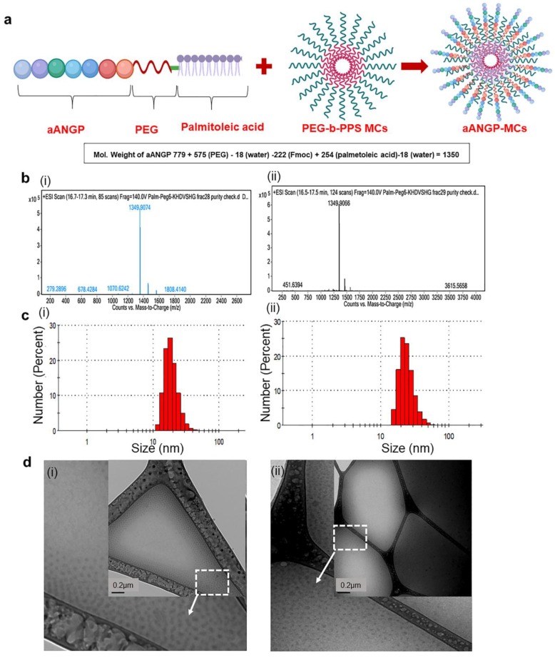
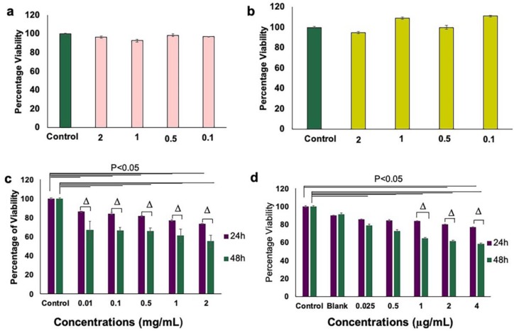
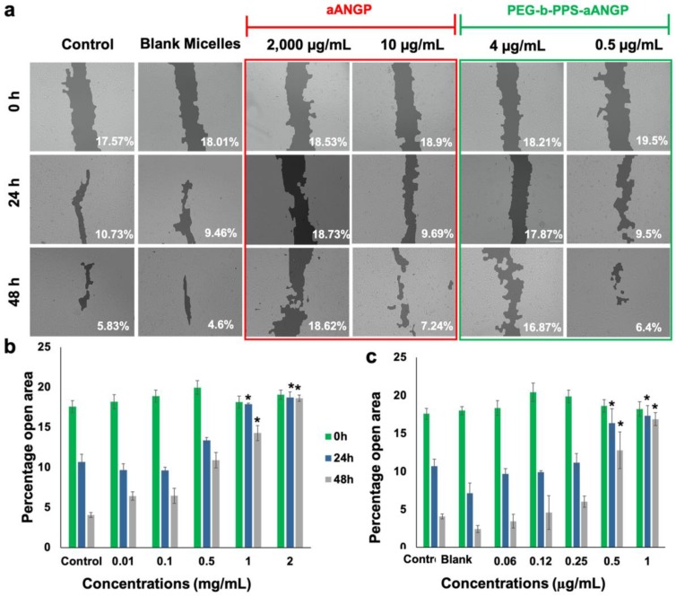
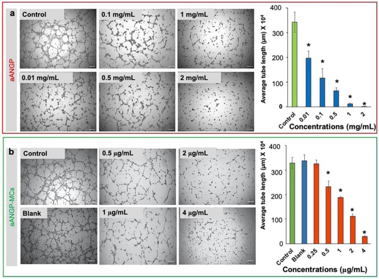
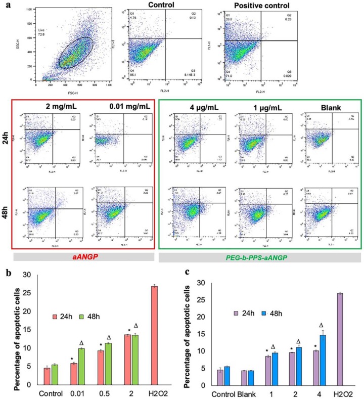
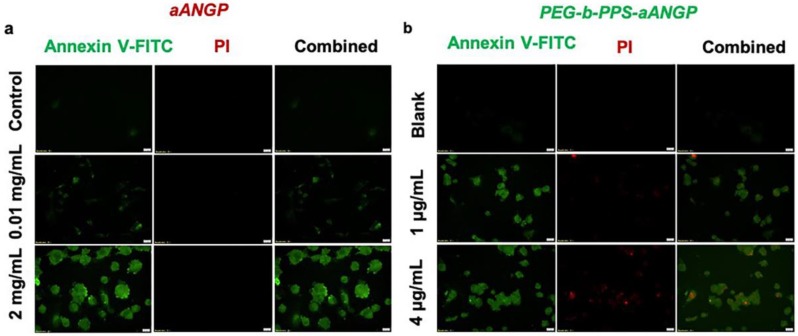
Diabetic retinopathy (DR), Retinopathy of Pre-maturity (ROP), and Age-related Macular Degeneration (AMD) are multifactorial manifestations associated with abnormal growth of blood vessels in the retina. These three diseases account for 5% of the total blindness and vision impairment in the US alone. The current treatment options involve heavily invasive techniques such as frequent intravitreal administration of anti-VEGF (vascular endothelial growth factor) antibodies, which pose serious risks of endophthalmitis, retinal detachment and a multitude of adverse effects stemming from the diverse physiological processes that involve VEGF. To overcome these limitations, this current study utilizes a micellar delivery vehicle (MC) decorated with an anti-angiogenic peptide (aANGP) that inhibits αvβ3 mediated neovascularization using primary endothelial cells (HUVEC). Stable incorporation of the peptide into the micelles (aANGP-MCs) for high valency surface display was achieved with a lipidated peptide construct. After 24 h of treatment, aANGP-MCs showed significantly higher inhibition of proliferation and migration compared to free from aANGP peptide. A tube formation assay clearly demonstrated a dose-dependent angiogenic inhibitory effect of aANGP-MCs with a maximum inhibition at 4 μg/mL, a 1000-fold lower concentration than that required for free from aANGP to display a biological effect. These results demonstrate valency-dependent enhancement in the therapeutic efficacy of a bioactive peptide following conjugation to nanoparticle surfaces and present a possible treatment alternative to anti-VEGF antibody therapy with decreased side effects and more versatile options for controlled delivery.

Keywords: Micelles; PEG-b-PPS; VEGF; anti-angiogenic; integrin.


Conflict of interest statement

The authors declare no conflict of interest.

Figures

Figure 1
Figure 1
Design and characterization of aANGP-micellar delivery vehicle (MCs). (a) Schematic representation of the aANGP lipidated peptide construct and its insertion into poly (ethylene glycol)-b-poly (propylene sulfide) (PEG-b-PPS) MCs. (b) Total ion chromatograms of the fractions collected at retention times of 42 (i) and 45 (ii) min showing the peak obtained for the modified aANGP. (c) Dynamic Light Scattering (DLS) spectrum obtained for (i) PEG-b-PPS (blank) micelles and (ii) aANGP-MCs with the average particle size of 20–50 nm. (d) Cryogenic-Transmission Electron Microscopy (Cryo-TEM) images of (i) PEG-b-PPS (Blank) micelles and (ii) aANGP-MCs (inset) showed their respective morphology and size to both be spherical and 20–50 nm in diameter. Outer image is manually enlarged and cropped section of the white square part of the inset image.
Figure 2
Figure 2
Immunostaining of angiogenic markers in Human Umbilical Vein Endothelial Cell line (HUVECs). Cells were stained with Platelet Endothelial Cell Adhesion Molecule-1 (PECAM-1) (green), vWF (red) and nucleus with DAPI (blue). The expression of PECAM-1 and von Willebrand Factor (VWF) is evident in HUVECs regardless of basic Fibroblast Growth Factor (bFGF) exposure.
Figure 3
Figure 3
Expression of αvβ3 by HUVECs under different conditions (a) Flow cytometry analysis demonstrated significant expression of integrin αvβ3 in overnight starved cells. (i) Side scatter (SSC) vs. forward scatter (FSC) plot used to gate live cells and resulting (ii) histograms from which αvβ3 expression was (iii) quantified for different culture conditions. Colors in (ii) match the conditions shown in the x-axis of (iii). Values were expressed as mean ± SD, n = 3. p < 0.005 was considered significant. (b) Immunostaining was performed by (i) staining HUVECs with anti-human CD51/CD61 antibody to detect integrin αvβ3 (green). Nuclei were stained by DAPI (blue). Integrin αvβ3 expression was highest under overnight starved conditions when compared to control and 2 h starvation. (ii) Quantification of the images in (i) obtained using Image J. Value was expressed as mean ± SD, n = 3. p < 0.05 was considered significant.
Figure 4
Figure 4
Comparison of HUVEC viability in the presence between aANGP-MCs and free form peptide as assessed by MTT assay. (a) Graph showing cell viability after treatment with aANGP without overnight starvation and (b) after treatment with aANGP with overnight starvation using 10 ng/mL of VEGF. (c) Viability results obtained after treatment with free form aANGP and (d) with aANGP-MCs after 24 and 48 h. Δ-Significance between the samples at 24 and 48 h. Values were expressed as mean ± SD, n = 3. Δ p < 0.05.
Figure 5
Figure 5
Effect of aANGP and aANGP-MCs on the migration of HUVECs. (a) HUVECs were treated with different concentrations of aANGP and aANGP-MCs for 24 and 48 h and imaged at 0, 24, and 48 h using an inverted microscope at 10× magnification. Bar graphs represent the quantification of the images obtained for (b) aANGP and (c) aANGP-MCs. Values were expressed as mean SD, n = 3. * p < 0.005.
Figure 6
Figure 6
Effect of aANGP and aANGP-MCs on HUVEC tube formation: HUVECs were seeded on.plates coated with ECM gel, incubated for 4 h with different concentrations of aANGP and aANGP-MCs and then imaged using an inverted microscope at 4× magnification. Images obtained for treatment with different concentrations of (a) free form aANGP and (b) aANGP-MCs. Bar graph represents the quantification of the tubes formed obtained using Image J. Values were expressed as mean ± SD, n = 3. * p < 0.0005.
Figure 7
Figure 7
Apoptosis induced by aANGP and aANGP-MCs: HUVECs were treated with different concentrations of aANGP and aANGP-MCs for 24 and 48 h followed by Annexin V-FITC and PI staining. (a) Dot plots obtained after analyzing aANGP treated HUVECs using Flow cytometry at 24 and 48 h. Bar graphs represent the quantification of the dot plots obtained for (b) aANGP and (c) aANGP-MCs treated HUVECs were stained with Annexin V-FITC and PI and imaged using a fluorescent microscope at 20× magnification. Values were expressed as mean ± SD, n = 3. * (24h) and Δ (48h) * p < 0.05.
Figure 8
Figure 8
Confocal images of apoptosis induced by aANGP and aANGP-MCs: (a) Images obtained for aANGP and (b) aANGP-MCs. Green—Annexin V- FITC (apoptosis), red—PI (necrosis). Values were expressed as mean ± SD, n = 3. p < 0.005.

References

    1. Tarr J.M., Kaul K., Chopra M., Kohner E.M., Chibber R. Pathophysiology of Diabetic Retinopathy. ISRN Ophthalmol. 2013;2013:343560. doi: 10.1155/2013/343560. - DOI - PMC - PubMed
    1. Retinopathy of Prematurity|National Eye Institute. [(accessed on 20 December 2019)]; Available online: https://www.nei.nih.gov/learn-about-eye-health/eye-conditions-and-diseas....
    1. Nowak J.Z. Age-related macular degeneration (AMD): Pathogenesis and therapy. Pharmacol. Rep. PR. 2006;58:353–363. - PubMed
    1. Brey E.M., McIntire L.V. 59—Vascular Assembly in Engineered and Natural Tissues. In: Atala A., Lanza R., Thomson J.A., Nerem R.M., editors. Principles of Regenerative Medicine. Academic Press; San Diego, CA, USA: 2008. pp. 1020–1037.
    1. Hennig R., Goepferich A. Nanoparticles for the treatment of ocular neovascularizations. Eur. J. Pharm. Biopharm. 2015;95:294–306. doi: 10.1016/j.ejpb.2015.02.027. - DOI - PubMed

LinkOut - more resources