. 2020 Jun;56(6):1405-1416.
doi: 10.3892/ijo.2020.5017. Epub 2020 Mar 19.

The resveratrol analogue, HS‑1793, enhances the effects of radiation therapy through the induction of anti‑tumor immunity in mammary tumor growth

Affiliations

The resveratrol analogue, HS‑1793, enhances the effects of radiation therapy through the induction of anti‑tumor immunity in mammary tumor growth

Joong Sun Kim et al. Int J Oncol. 2020 Jun.

Abstract

Radiotherapy can induce the infiltration of immune suppressive cells which are involved in promoting tumor progression and recurrence. A number of natural products with immunomodulating abilities have been gaining attention as complementary cancer treatments. This attention is partly due to therapeutic strategies which have proven to be ineffective as a result of tumor‑induced immunosuppressive cells found in the tumor microenvironment. The present study investigated whether HS‑1793, a resveratrol analogue, can enhance the antitumor effects by inhibiting lymphocyte damage and immune suppression by regulatory T cells (Tregs) and tumor‑associated macrophages (TAMs), during radiation therapy. FM3A cells were used to determine the role of HS‑1793 in the radiation‑induced tumor immunity of murine breast cancer. HS‑1793 treatment with radiation significantly increased lymphocyte proliferation with concanavalin A (Con A) stimulation and reduced the DNA damage of lymphocytes in irradiated tumor‑bearing mice. The administration of HS‑1793 also decreased the number of Tregs, and reduced interleukin (IL)‑10 and transforming growth factor (TGF)‑β secretion in irradiated tumor‑bearing mice. In addition, HS‑1793 treatment inhibited CD206+ TAM infiltration in tumor tissue when compared to the controls or irradiation alone. Mechanistically, HS‑1793 suppressed tumor growth via the activation of effector T cells in irradiated mice. On the whole, the findings of the present study reveal that HS‑1793 treatment improves the outcome of radiation therapy by enhancing antitumor immunity. Indeed, HS‑1793 appears to be a good therapeutic candidate for use in combination with radiotherapy in breast cancer.

Keywords: radiotherapy; HS‑1793; resveratrol analogue; regulatory T cells; tumor‑associated macrophages; antitumor immunity; breast cancer.


Figures

Figure 1
Figure 1
Effect of HS-1793 on the viability in 137Cs γ-radiation-exposed FM3A cells. (A) Structure of the resveratrol analogue, HS-1793. (B) FM3A cells were treated with the indicated concentrations of HS-1793 (0.31-10 µg/ml) or (C) radiation doses (0-16 Gy) for 24 and 48 h. (D) FM3A cells were treated with the indicated concentrations of HS-1793 for 24 h pior to 4 Gy irradiation. The cells were then cultured for 24 h and cell viability was determined by MTT assay. #P<0.05 as compared with the control and *P<0.05 as compared with irradiation alone (IR); one-way analysis of variance with Tukey's post hoc test. (E) FM3A cells were treated with the indicated concentrations of HS-1793 at 37°C for 24 h prior to irradiation (2, 4, 6, 8, 10 Gy) for clonogenic assays. After approximately 14 days, colonies were counted. Each value in treated cells was calculated with respect to the untreated control. All samples were performed in triplicate and experiments were repeated three times. Results are expressed as percentages of control, and data are the means ± standard deviations of 3 independent experiments. *P<0.05 as compared with irradiation alone (IR); one-way analysis of variance with Dunnett's post hoc test. (F) Representative images of clonogenic assays showing that radiation inhibited the colony forming capacity of cancer cells in a dose-dependent manner and that HS-1793 enhanced the tumor suppressive effect of radiation.
Figure 2
Figure 2
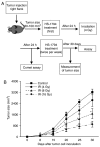
Optimal dose of radiation for combined treatment of HS-1793 in tumor-bearing mice. (A) Schematic diagram showing the schedule of combination treatments in FM3A tumor-bearing mice. (B) For the evaluation of the optimal dose of radiation for combination treatment with HS-1793, FM3A cells (2×10⁶ cells/50 µl) were inoculated subcutaneously into the right flanks of female C3H/He mice. When the tumor grew to a size of approximately 80-125 mm3, the mice were irradiated with various doses (4, 8 and 16 Gy) and tumor size was assessed once a week for 30 days. Data reported as the mean tumor size ± SD of 5 mice per group. *P<0.05 as compared with the control; one-way analysis of variance with Dunnett's post hoc test.
Figure 3
Figure 3
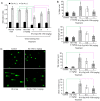
Effect of HS-1793 on the proliferation and DNA damage of radiation induced lymphocytes in tumor-bearing mice. FM3A cells (2×10⁶ cells/50 µl) were inoculated subcutaneously into the right flanks of female C3H/He mice. At a tumor size of approximately 80-100 mm3, the mice were treated with intraperitoneal injections of HS-1793 (0.5 and 1 mg/kg) and were irradiated with 4 Gy. After 24 h, the mice were treated with HS-1793 twice a week for 3 weeks. (A) Con A-stimulated lymphocyte proliferation assay was performed with splenocytes isolated from each group at 3 weeks after the HS-1793 injection, and proliferation was measured on day 3 by the incorporation of BrdU during the final 24 h. (B) Comet assays were performed with splenocytes isolated from each group at 24 h after irradiation. The representative comet parameters (% DNA in tail, tail length, tail moment, and olive tail moment) and (C) the photomicrographs of comet length are presented for each condition. A minimum of 100 cells were analyzed using Metafer 4 software. Data reported as the means ± SD from 5 mice per group. #P<0.05 as compared with the control and *P<0.05 as compared with irradiation alone (IR); one-way analysis of variance with Tukey's post hoc test.
Figure 4
Figure 4
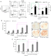
Effect of HS-1793 on Tregs of irradiated tumor-bearing mice. After the final injections of HS-1793, splenocytes from aseptically removed spleen and paraffin-embedded sections of tumor tissue were prepared. (A) Splenocytes were cultured with concanavalin A (5 µg/ml) for 24 h and the cultured supernatants were analyzed for cytokine concentrations by ELISA. (B) Splenocytes were analyzed for the expression of CD4, CD25 and intracellular FoxP3 by flow cytometry. The percent gated cells of FoxP3+CD25+ cells among CD4+ T cells is shown. (C) Immunofluorescence staining (×400) for CD25+ T cell infiltration in tumor tissue. Data reported as the means ± SD from 5 mice per group. #P<0.05 as compared with the control and *P<0.05 as compared with irradiation alone (IR); one-way analysis of variance with Tukey's post hoc test.
Figure 5
Figure 5
Effect of HS-1793 on IFN-γ expressing CD8+ T cells of irradiated tumor-bearing mice. After the final injections of HS-1793, splenocytes were analyzed for the expression of CD8 and intracellular IFN-γ by flow cytometer and ELISPOT assay. (A) Percentage gated cells of CD8+ T cells and IFN-γ producing CD8+ T cells were calculated. (B) Splenocytes (5×10⁴ cells/well) were seeded in ImmunoSpot plates coated with anti-mouse IFN-γ antibody and incubated with stimulant cocktail. ImmunoSpot assay used to show the number of spot and the representative images in each condition. (C) Splenocytes were cultured with concanavalin A (5 µg/ml) for 24 h and the amounts of IFN-γ in culture supernatants were determined by ELISA. Data reported as the means ± SD from 5 mice per group. #P<0.05 or ##P<0.05 as compared with the control and *P<0.05 as compared with irradiation alone (IR); one-way analysis of variance with Tukey's post hoc test.
Figure 6
Figure 6
Effect of HS-1793 on infiltration of tumor-associated macrophages (TAMs) in irradiated tumor-bearing mice. Paraffin-embedded sections (5-µm-thick) were prepared from tumor tissue and stained for CD206 and IFN-γ with immunofluorescent antibodies. Immunofluorescence staining (×200) shown for IFN-γ deposition and CD206+ macrophages.
Figure 7
Figure 7
Anti-tumor activity of HS-1793 in irradiated tumor bearing mice. FM3A cells (2×10⁶ cells/50 µl) were inoculated subcutaneously on the right flank of female C3H/He mice. At a tumor size of 80-125 mm3, the mice were treated with intraperitoneal injections of HS-1793 (0.5 and 1 mg/kg) and were irradiated with 4 Gy. After 24 h, the mice were treated with HS-1793 twice a week for 3 weeks. (A) The cytotoxicity of splenocytes against target FM3A tumor cells was measured with LDH release assaying kit. (B) For the evaluation of combination effects of HS-1793 and radiation, a single inoculation of tumor cells was performed simultaneously with intraperitoneal injections of HS-1793 twice a week after irradiation for 3 weeks. Tumor size was assessed weekly. Data reported as the means ± SD from 5 mice per group. (C) Representative photographs of mouse with tumor in each group are shown. #P<0.05 as compared with the control and *P<0.05 as compared with irradiation alone (IR); one-way analysis of variance with Tukey's post hoc test.

Similar articles

Cited by

References

    1. Basu P, Maier C. Phytoestrogens and breast cancer: In vitro anticancer activities of isoflavones, lignans, coumestans, stilbenes and their analogs and derivatives. Biomed Pharmacother. 2018;107:1648–1666. doi: 10.1016/j.biopha.2018.08.100. - DOI - PubMed
    1. Clarke M, Collins R, Darby S, Davies C, Elphinstone P, Evans V, Godwin J, Gray R, Hicks C, James S, et al. Effects of radio-therapy and of differences in the extent of surgery for early breast cancer on local recurrence and 15-year survival: An overview of the randomised trials. Lancet. 2005;366:2087–2106. doi: 10.1016/S0140-6736(05)67887-7. - DOI - PubMed
    1. Turashvili G, Brog E. Tumor heterogeneity in breast cancer. Front Med (Lausanne) 2017;4:227. doi: 10.3389/fmed.2017.00227. - DOI - PMC - PubMed
    1. Grantzau T, Overgaard J. Risk of second non-breast cancer after radiotherapy for breast cancer: A systematic review and meta-analysis of 762,468 patients. Radiother Oncol. 2015;114:56–65. doi: 10.1016/j.radonc.2014.10.004. - DOI - PubMed
    1. Bartelink H, Horiot JC, Poortmans PM, Struikmans H, Van den Bogaert W, Fourquet A, Jager JJ, Hoogenraad WJ, Oei SB, Wárlám-Rodenhuis CC, et al. Impact of a higher radiation dose on local control and survival in breast-conserving therapy of early breast cancer: 10-Year results of the randomized boost versus no boost EORTC 22881-10882 trial. J Clin Oncol. 2007;25:3259–3265. doi: 10.1200/JCO.2007.11.4991. - DOI - PubMed

MeSH terms