. 2020 Jun;139(6):1089-1104.
doi: 10.1007/s00401-020-02150-w. Epub 2020 Mar 31.

Intracellular calcium leak as a therapeutic target for RYR1-related myopathies

Affiliations

Intracellular calcium leak as a therapeutic target for RYR1-related myopathies

Alexander Kushnir et al. Acta Neuropathol. 2020 Jun.

Abstract

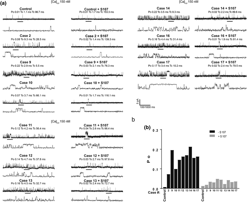
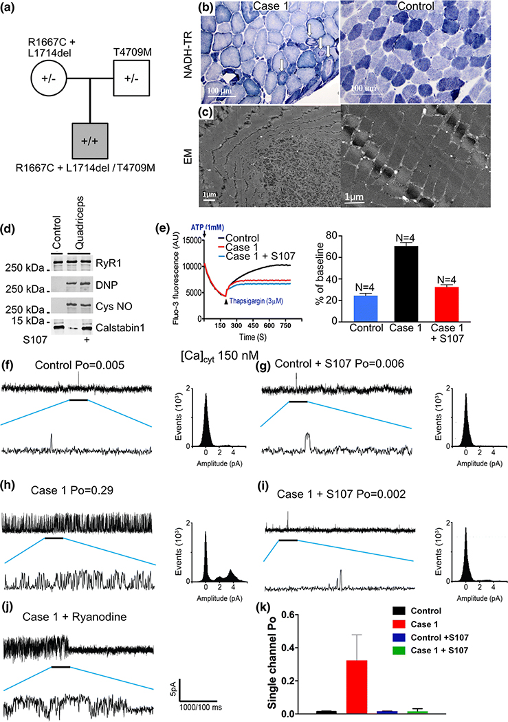
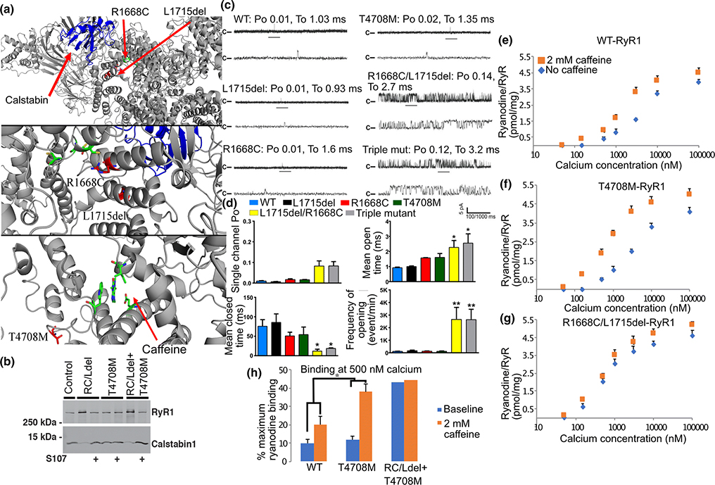
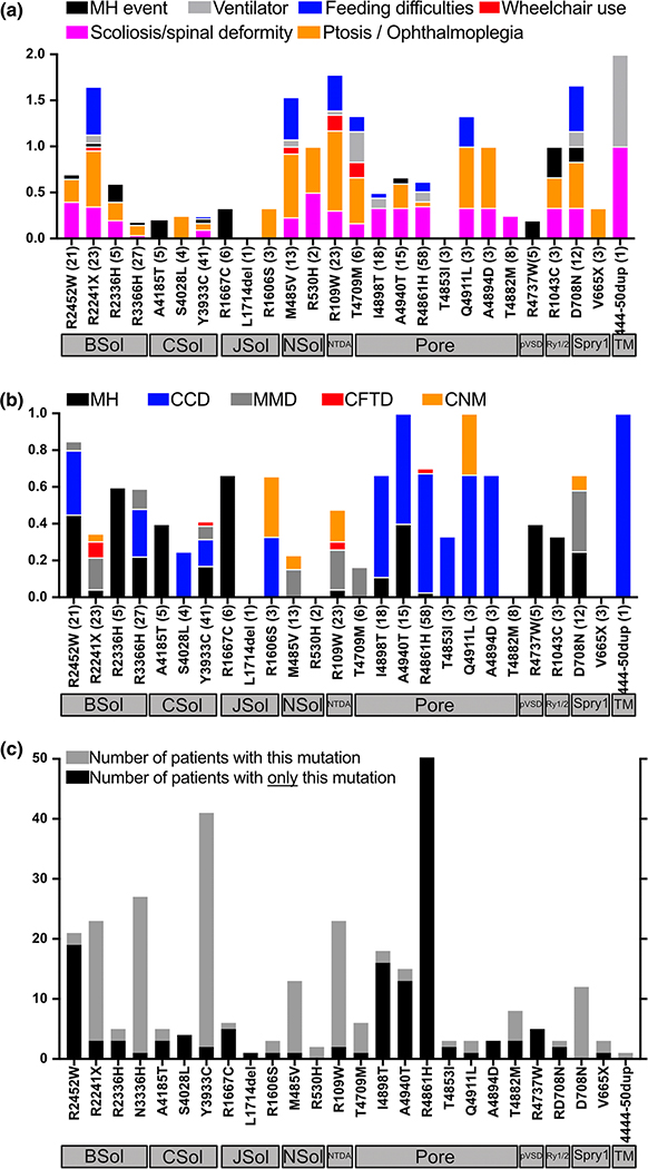
RYR1 encodes the type 1 ryanodine receptor, an intracellular calcium release channel (RyR1) on the skeletal muscle sarcoplasmic reticulum (SR). Pathogenic RYR1 variations can destabilize RyR1 leading to calcium leak causing oxidative overload and myopathy. However, the effect of RyR1 leak has not been established in individuals with RYR1-related myopathies (RYR1-RM), a broad spectrum of rare neuromuscular disorders. We sought to determine whether RYR1-RM affected individuals exhibit pathologic, leaky RyR1 and whether variant location in the channel structure can predict pathogenicity. Skeletal muscle biopsies were obtained from 17 individuals with RYR1-RM. Mutant RyR1 from these individuals exhibited pathologic SR calcium leak and increased activity of calcium-activated proteases. The increased calcium leak and protease activity were normalized by ex-vivo treatment with S107, a RyR stabilizing Rycal molecule. Using the cryo-EM structure of RyR1 and a new dataset of > 2200 suspected RYR1-RM affected individuals we developed a method for assigning pathogenicity probabilities to RYR1 variants based on 3D co-localization of known pathogenic variants. This study provides the rationale for a clinical trial testing Rycals in RYR1-RM affected individuals and introduces a predictive tool for investigating the pathogenicity of RYR1 variants of uncertain significance.

Keywords: Calcium; Central core disease; Genetics; RyR1-related myopathy; Ryanodine receptor; Therapeutics.


Conflict of interest statement

Conflict of interest statement: Columbia University and ARM own stock in ARMGO, Inc. a company developing compounds targeting RyR and have patents on Rycals.

Figures

Figure 1
Figure 1. Reduced calstabin1 binding to RYR1-RM channels restored by ex-vivo treatment with Rycal.
(a) RyR1 was immunoprecipitated using a RyR1-specific antibody from 0.1 mg of skeletal muscle lysates obtained from RYR1-RM affected individuals included in this study in the absence and presence of 1.0 μM S107. Co-immunoprecipitated calstabin1 is assessed using RyR antibody and calstabin specific antibodies. Oxidation of RyR1 is determined by first derivatizing the carbonyl groups in the protein side chains in the immunoprecipitate with dinitrophenylhydrazone (DNP, Abcam, 178020) and then detecting the DNP signal associated with RyR using an anti-DNP antibody. See Table 1 for RYR1 variants. (b) Quantification of immunoblot data. Control Calstabin/RyR1 set to 4/1. * p<0.01 compared to control, ** p<0.01 S107 treated vs. baseline lysates. (c). Calpain activity of muscle lysates determined using a commercial kit (Abcam). The calpain activity assay protocol is based on the detection of cleavage of calpain substrate and the relative change in fluorescence signal/μg lysate after 1 h at 37°C is plotted (control arbitrarily set to 1.0).
Figure 2
Figure 2. Increased calcium leak in single RYR1-RM channels reconstituted in planar lipid bilayer.
SR microsomes containing single RyR1 channels isolated from individuals with RYR1-RM fused with planar lipid bilayer. Channel opening events are recorded as an upward deflection. Area on upper graph is expanded as the lower graph. Po = opening probability, To = time open, Tc = time closed. (a) Single-channel recordings of RyR1 from muscle lysates that were either treated or untreated with S107 (1.0 μM) from control and RYR1-RM affected individuals. Recordings were performed at 150 nM Ca2+. (b), Bar graph summarizing single-channel Po. N=3 per group. Limited availability of skeletal muscle precluded analyses in Cases 3–8 and 15.
Figure 3
Figure 3. Ca2+ leak from RYR1-RM SR microsomes.
(a) SR Ca2+ leak measured in microsomes (5μg/mL) from RYR1-RM muscle lysates. The Ca2+ leak was compared for control, RYR1-RM, and RYR1-RM treated with 1 uM S107 as indicated. Control data is shown in the first panel in blue and is represented by dotted line (....) in remaining traces for comparison. Inserts above traces show magnification of first 20 s after addition of thapsigargin. Bar graphs represent the quantification of the increase in Fluo-4/s over the first 5 seconds after addition of thapsigargin. (b) Quantification of Ca2+ leak experiments. N=3 in each group. Mutant channels at baseline exhibit continuous leak which does not occur in control channels or in mutant channels treated with S107. Limited availability of skeletal muscle precluded analyses in Cases 3–8 and 15.
Figure 4
Figure 4. Genotype-structure-function analysis of Case 1.
(a) Inheritance pattern of RYR1 variations. (b) NADH-TR stain and (c) electron microscopic of quadriceps muscle biopsy from Case 1 alongside control showing cores (arrows) of quadriceps muscle exhibiting a discrete area of myofibrillar derangement and paucity of mitochondria in affected myofibers. (d) RyR1 immunoprecipitated from muscle biopsies from control and RYR1-RM-affected muscle (quadriceps) comparing oxidation, nitrosylation, and calstabin1/RyR1 association (with and without Rycal S107 treatment). (e) Ca2+ leak assay in the presence and absence of Rycal S107 with bar graph, *P<0.05. (f) Single-channel data from normal control human quadriceps muscles in the absence and (g) presence of Rycal S107. (h) Single-channel data from Case 1’s quadriceps muscle in the absence (I) and presence of Rycal S107. There are multiple partial openings or subconductance states evident in the channels recorded from Case 1 in the absence of Rycal S107 which are seen in calstabin1 depleted channels that exhibit defective closing (leak). These subconductance states are not observed in the Rycal S107 treated muscle consistent with repair of channel leak. Scale bar represents current amplitude and time scale for compressed and expanded tracings. Amplitude histograms are shown for each experiment. (j) Channel from Case 1 treated with ryanodine which locks the channel in a sub-conductance state confirming the identity of the channel. (k) Bar graph shows quantification of single-channel Po data, N=5 per group, Tracings show samples of representative channel behavior in the bilayer. All analyses were performed on a minimum of two minutes of channel recording.
Figure 5
Figure 5. Characterization of defective RyR1 channel function Case 1.
(a) Localization of maternal variants (RYR1-R1668C, L1715del – numbering based on rabbit RyR1) from Case 1 to near the calstabin1 (yellow) binding site in RyR1 (top and middle panel). Localization of paternal variant (RYR1-T4708M) near the caffeine binding site (green). (b) Recombinant RYR1 variants (RC/Ldel: RYR1-R1668C, L1715del; T4708M: RYR1-T4708M and the combination of all three mutations: RC/Ldel on one cDNA and T4708M on another) immunoprecipitated from transfected HEK cell lysates and immunoblotted for calstabin in the presence or absence of Rycal S107. The RC/Ldel but not the T4708M result in decreased binding of calstabin1 to RyR1 that is restored by Rycal S107. (c) Single-channel current recordings from WT and mutant recombinant RyR1 at 150 nM [Ca2+]cis. (d) Bar graph shows quantification of data. N=6 per group, (* P<0.05). (e) 3H-ryanodine binding to recombinant RyR1 lysates expressing: (e) WT, (f) paternal, or (g) maternal variants, in the presence or absence of 2 mM caffeine and at the indicated [Ca2+]. (h) Bar graph shows quantification of 3H-ryanodine binding data at 500 nM [Ca2+]. *, P<0.05.
Figure 6
Figure 6. RYR1 dataset and localization of variants to the 3D RyR1 structure.
(A) Summary of variants in gnomAD database and RYR1 dataset with amino acid position on the X-axis. Channel structural domains highlighted based on references [9,47]. Each report of a variant in the RYR1 dataset is represented by a grey dot. Each variant reported in gnomAD is represented by a black dot. Inverted black triangles represent gnomAD variants with a frequency >0.01%. Inverted grey triangles represent variants included in this study. (B) Top - A single RyR1 protomer with locations of calstabin1 (yellow), the SPRY (cyan), Bsol (green), and pore (orange) domains highlighted. Bottom – Close-up of SPRY domain (cyan) with variants (red) present in the RYR1 dataset and their co-localization with calstabin1 (yellow). (C) Overview of the homotetrameric RyR1 channel. Variants in the Bsol domain are highlighted in red.
Figure 7
Figure 7. Analysis of RYR1 variants examined in the present study:
(A) Variants from individuals included in this study are on the X-axis. Number of individuals in the RYR1 dataset with that variant in parentheses. Variants are ordered based on their structural domain localization on the channel as depicted by the horizontal grey boxes under the variants. Y-axis represents the percentage of individuals with a specific symptom. For example, amongst 12 entries of D708N variant (located in the SPRY1 domain) in the RYR1 dataset: 35% have scoliosis; 40% have ptosis; 40% have feeding difficulties, 8% are wheelchair-bound, and 8% had an MH event. (B) Y-axis represents the percentage of individuals with a specific histopathologic finding (and/or laboratory diagnosis of MH who may or may not have had a clinical event). For example, for the 12 entries of D708N variant in RYR1 dataset, 25% have a laboratory diagnosis of MH, 40% have MmD, 8% of entries have CNM. (C) Y-axis grey bar is the total number of times each RYR1 variant appears in the RYR1 dataset. Y-axis black bar is the number of times a variant appears in the RYR1 dataset without additional variants.

References

    1. Amburgey K, McNamara N, Bennett LR, McCormick ME, Acsadi G, Dowling JJ (2011) Prevalence of congenital myopathies in a representative pediatric united states population. Ann Neurol 70:662–665. doi:10.1002/ana.22510 - DOI - PubMed
    1. Andersson DC, Betzenhauser MJ, Reiken S, Meli AC, Umanskaya A, Xie W, Shiomi T, Zalk R, Lacampagne A, Marks AR (2011) Ryanodine Receptor Oxidation Causes Intracellular Calcium Leak and Muscle Weakness in Aging. Cell metabolism 14:196–207. doi:10.1016/j.cmet.2011.05.014 - DOI - PMC - PubMed
    1. Andersson DC, Meli AC, Reiken S, Betzenhauser MJ, Umanskaya A, Shiomi T, D’Armiento J, Marks AR (2012) Leaky ryanodine receptors in beta-sarcoglycan deficient mice: a potential common defect in muscular dystrophy. Skelet Muscle 2:9. doi:10.1186/2044-5040-2-9 - DOI - PMC - PubMed
    1. Avila G, O’Brien JJ, Dirksen RT (2001) Excitation--contraction uncoupling by a human central core disease mutation in the ryanodine receptor. Proc Natl Acad Sci U S A 98:4215–4220. doi:10.1073/pnas.071048198 - DOI - PMC - PubMed
    1. Bellinger AM, Reiken S, Carlson C, Mongillo M, Liu X, Rothman L, Matecki S, Lacampagne A, Marks AR (2009) Hypernitrosylated ryanodine receptor calcium release channels are leaky in dystrophic muscle. Nat Med 15:325–330. doi:10.1038/nm.1916 - DOI - PMC - PubMed

Publication types

MeSH terms

Substances