. 2021 Apr;39(7):2617-2627.
doi: 10.1080/07391102.2020.1751300. Epub 2020 Apr 15.

In-silico approaches to detect inhibitors of the human severe acute respiratory syndrome coronavirus envelope protein ion channel

Affiliations

In-silico approaches to detect inhibitors of the human severe acute respiratory syndrome coronavirus envelope protein ion channel

Manoj Kumar Gupta et al. J Biomol Struct Dyn. 2021 Apr.

Abstract

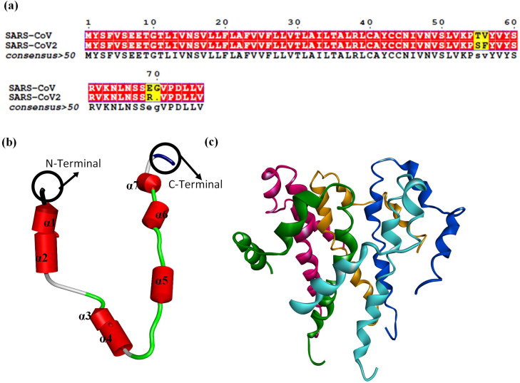
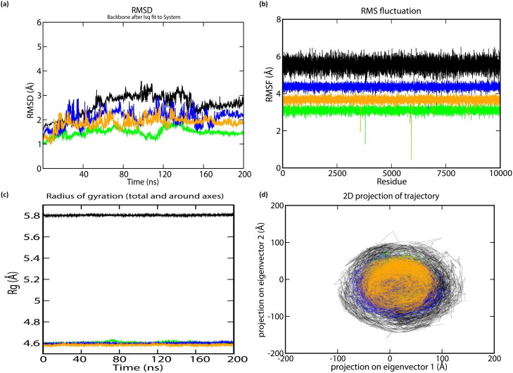
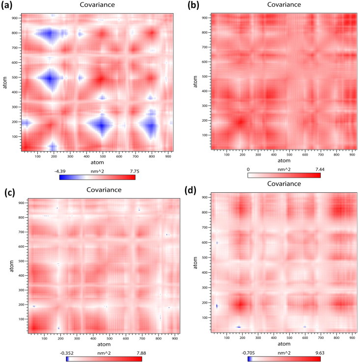
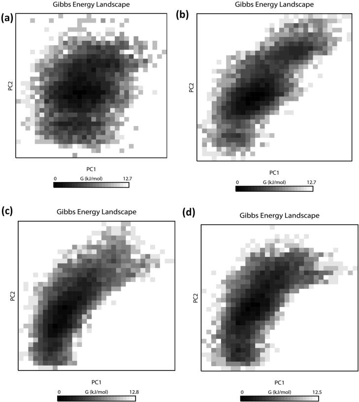
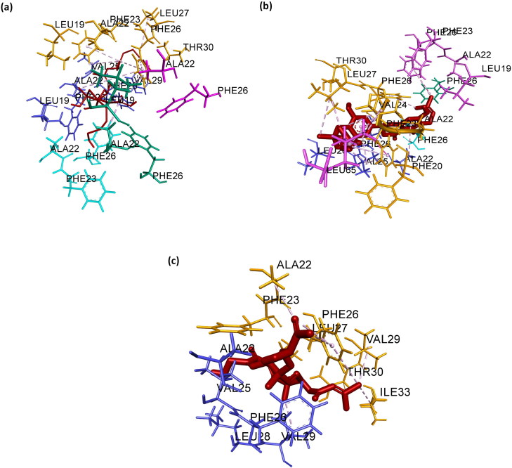
Recent outbreak of Coronavirus disease (COVID-19) pandemic around the world is associated with 'severe acute respiratory syndrome' (SARS-CoV2) in humans. SARS-CoV2 is an enveloped virus and E proteins present in them are reported to form ion channels, which is mainly associated with pathogenesis. Thus, there is always a quest to inhibit these ion channels, which in turn may help in controlling diseases caused by SARS-CoV2 in humans. Considering this, in the present study, authors employed computational approaches for studying the structure as well as function of the human 'SARS-CoV2 E' protein as well as its interaction with various phytochemicals. Result obtained revealed that α-helix and loops present in this protein experience random movement under optimal condition, which in turn modulate ion channel activity; thereby aiding the pathogenesis caused via SARS-CoV2 in human and other vertebrates. However, after binding with Belachinal, Macaflavanone E, and Vibsanol B, the random motion of the human 'SARS-CoV2 E' protein gets reduced, this, in turn, inhibits the function of the 'SARS-CoV2 E' protein. It is pertinent to note that two amino acids, namely VAL25 and PHE26, play a key role while interacting with these three phytochemicals. As these three phytochemicals, namely, Belachinal, Macaflavanone E & Vibsanol B, have passed the ADMET (Absorption, Distribution, Metabolism, Excretion and Toxicity) property as well as 'Lipinski's Rule of 5s', they may be utilized as drugs in controlling disease caused via SARS-COV2, after further investigation.Communicated by Ramaswamy H. Sarma.

Keywords: Coronaviruses; docking; modeling; molecular dynamics; severe acute respiratory syndrome.


Figures

Figure 1.
Figure 1.
(a) Sequence similarity between protein sequence of ‘SARS-CoV E’ and ‘SARS-CoV2 E’ protein. 3D structure of ‘SARS-CoV2 E’ protein, (b) One unit is composed of only seven α-helices and 8 loops. (c) Homopentamer. In (c), green, pink, orange, blue and cyan depicts chain A, B, C, D and E of the ‘SARS-CoV2 E’ pentameric protein.
Figure 2.
Figure 2.
The stability parameters for ‘SARS-CoV E’ protein during 200 ns: (a) RMSD of C-α (b) RMSF of C-α (c) Radius of gyration of C-α, and (d) Principle Component Analysis (PCA) of α-helix and β-strand movement. The trajectory projected to the two-dimensional space. Black, light green, blue, and orange lines represent ‘SARS-CoV2 E’ protein and Complexes A, B & C during 200 ns, respectively.
Figure 3.
Figure 3.
Comparative study of cross-correlation matrices of C-α atoms of modeled (a) ‘SARS-CoV2 E’ protein (b) Complex A (c) Complex B and (d) Complex C during 200 ns simulation. The range of motion indicated by various colors in the panel. Red indicates a positive correlation, whereas blue indicates anti-correlation.
Figure 4.
Figure 4.
Projections of the free energy landscape of (a) ‘SARS-CoV2 E’ protein (b) Complex A (c) Complex B and (d) Complex C during 200 ns simulation. Various colors in the panel indicate the range of motion, where dark black indicates the lowest energy configuration, and white shows the highest energy configuration.
Figure 5.
Figure 5.
‘Three-dimensional’ representation of intermolecular interaction in (a) Complex A, (b) Complex B and (c) Complex C. Green, pink, orange, blue and cyan depicts chain A, B, C, D and E of the pentameric ‘SARS-CoV2 E’ protein, respectively. Dark brown depicts ligand.

References

    1. Abraham M. J., Murtola T., Schulz R., Páll S., Smith J. C., Hess B., & Lindahl E. (2015). GROMACS: High performance molecular simulations through multi-level parallelism from laptops to supercomputers. SoftwareX, 1-2, 19–25. doi:10.1016/j.softx.2015.06.001 - DOI
    1. Altschul S. F., Gish W., Miller W., Myers E. W., & Lipman D. J. (1990). Basic local alignment search tool. Journal of Molecular Biology, 215(3), 403–410. doi:10.1016/S0022-2836(05)80360-2 - DOI - PubMed
    1. Ashour H. M., Elkhatib W. F., Rahman M. M., & Elshabrawy H. A. (2020). Insights into the recent 2019 novel coronavirus (SARS-CoV-2) in light of past human coronavirus outbreaks. Pathogens, 9(3), 186. doi:10.3390/pathogens9030186 - DOI - PMC - PubMed
    1. Berger O., Edholm O., & Jähnig F. (1997). Molecular dynamics simulations of a fluid bilayer of dipalmitoylphosphatidylcholine at full hydration, constant pressure, and constant temperature. Biophysical Journal, 72(5), 2002–2013. doi:10.1016/S0006-3495(97)78845-3 - DOI - PMC - PubMed
    1. Bertrand Teponno R., Kusari S., & Spiteller M. (2016). Recent advances in research on lignans and neolignans. Natural Product Reports, 33(9), 1044–1092. doi:10.1039/C6NP00021E - DOI - PubMed