. 2020 Apr 1;17(1):27.
doi: 10.1186/s12987-020-00188-2.

Angiomodulin (IGFBP7) is a cerebral specific angiocrine factor, but is probably not a blood-brain barrier inducer

Affiliations

Angiomodulin (IGFBP7) is a cerebral specific angiocrine factor, but is probably not a blood-brain barrier inducer

Ofri Bar et al. Fluids Barriers CNS. .

Abstract

Background: Several secreted factors have been identified as drivers of cerebral vasculature development and inducers of blood-brain barrier (BBB) differentiation. Vascular endothelial growth factor A (VEGF-A) is central for driving cerebral angiogenesis and Wnt family factors (Wnt7a, Wnt7b and norrin) are central for induction and maintenance of barrier properties. Expressed by developing neural tissue (neuron and glia progenitors), they influence the formation of central nervous system (CNS) vascular networks. Another type of factors are tissue-specific paracrine factors produced by endothelial cells (ECs), also known as 'angiocrine' factors, that provide instructive signals to regulate homeostatic and regenerative processes. Very little is known about CNS angiocrine factors and their role in BBB development. Angiomodulin (AGM) was reported to be expressed by developing vasculature and by pathological tumor vasculature. Here we investigated AGM in the developing CNS and its function as a potential BBB inducer.

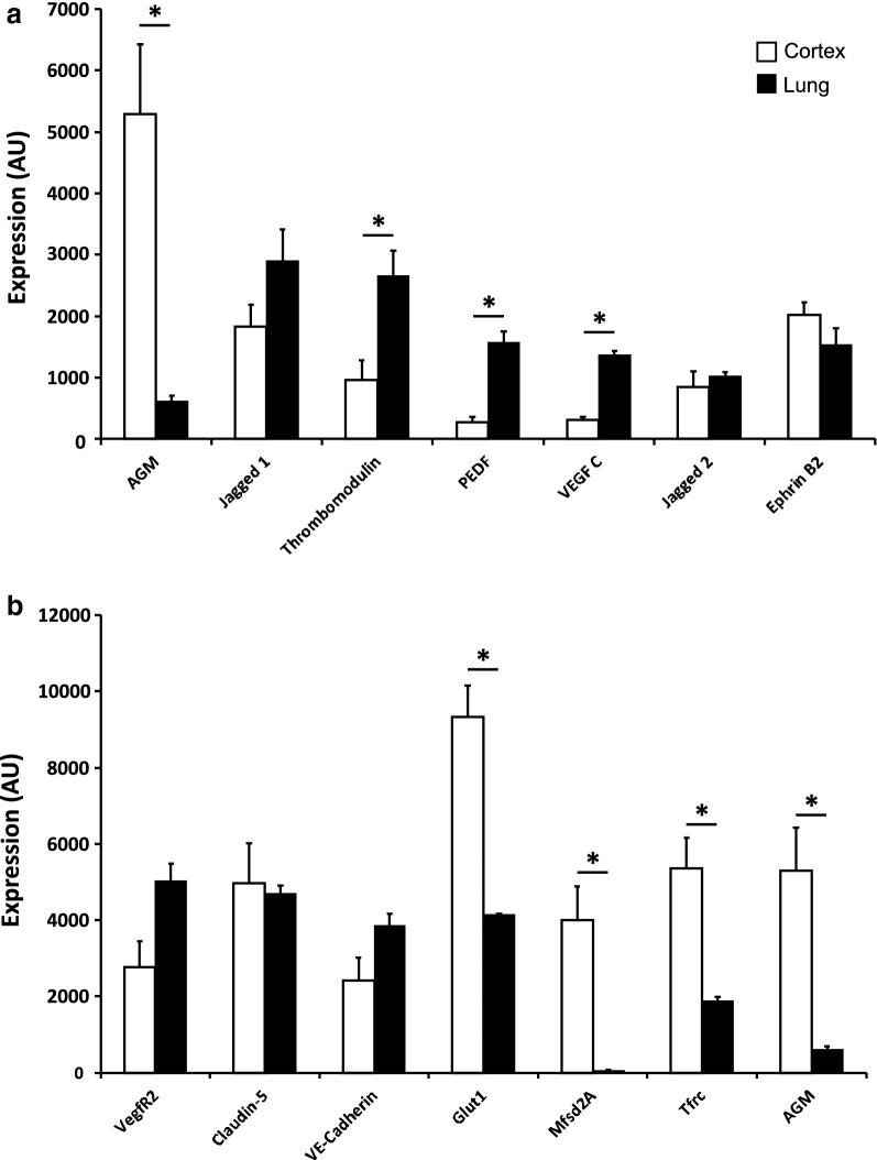
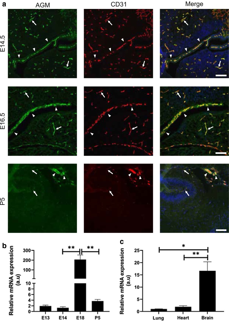
Methods: We analyzed microarray data to identify potential angiocrine factors specifically expressed at early stages of barrier formation. We then tested AGM expression with immunofluorescence and real-time PCR in various organs during development, post-natal and in adults. Permeability induction with recombinant proteins (Miles assay) was used to test potential interaction of AGM with VEGF-A.

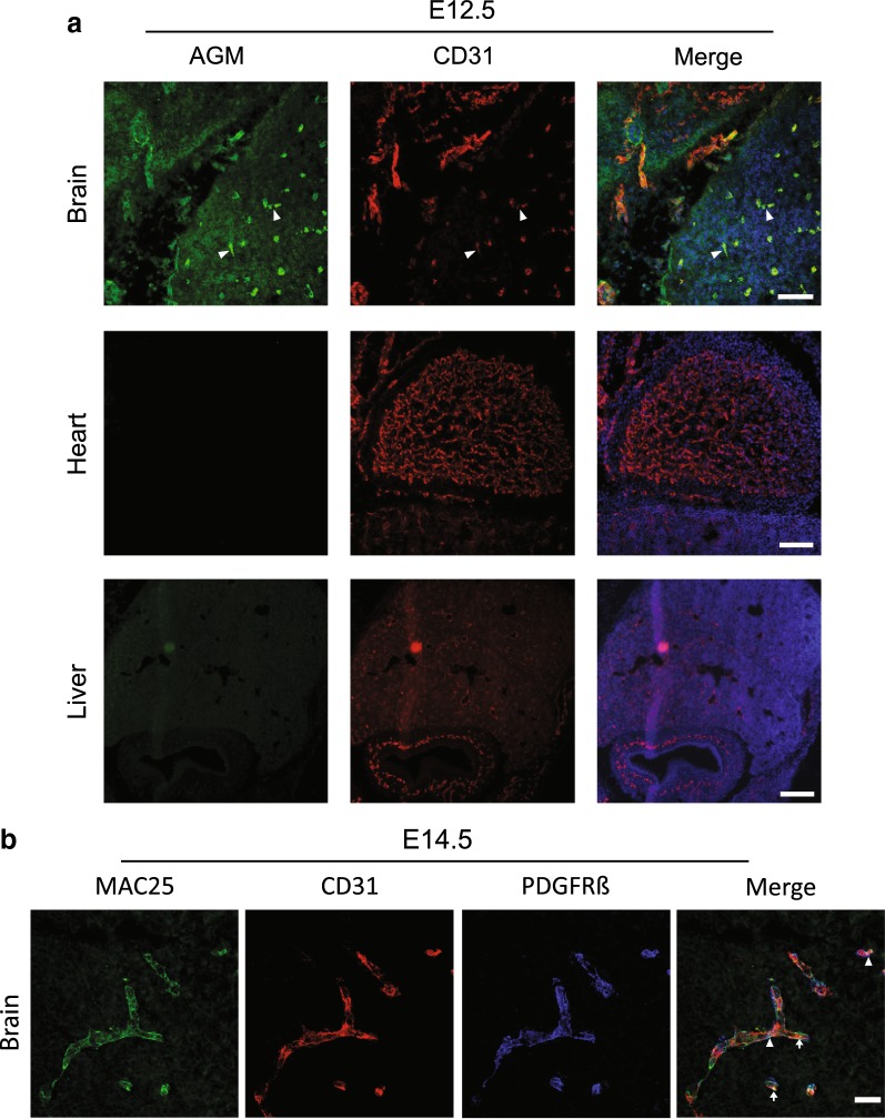
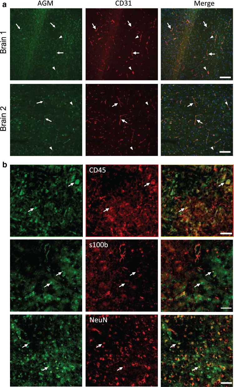
Results: Several angiocrine factors are differentially expressed by CNS ECs and AGM is a prominent CNS-specific angiocrine candidate. Contrary to previous reports, we found that AGM protein expression is specific to developing CNS endothelium and not to highly angiogenic developing vasculature in general. In skin vasculature we found that AGM antagonizes VEGF-A-induced vascular hyperpermeability. Finally, CNS AGM expression is not specific to BBB vasculature and AGM is highly expressed in non-BBB choroid-plexus vasculature.

Conclusions: We propose AGM as a developmental CNS vascular-specific marker. AGM is not a pan-endothelial marker, nor a general marker for developing angiogenic vasculature. Thus, AGM induction in the developing CNS might be distinct from its induction in pathology. While AGM is able to antagonize VEGF-A-induced vascular hyperpermeability in the skin, its high expression levels in non-BBB CNS vasculature does not support its potential role as a BBB inducer. Further investigation including loss-of-function approaches might elucidate AGM function in the developing CNS.

Keywords: Angiocrine factors; Angiomodulin (IGFBP7); Blood–brain barrier (BBB); Choroid plexus; Development; VEGF-A.


Conflict of interest statement

The authors declare that they have no competing interests.

Figures

Fig. 1
Fig. 1
Angiocrine factors are differentially expressed by cerebral endothelium at early stages of BBB induction and differentiation. Microarray data analysis from our previous study [4] comparing transcriptomes of lung and cortical endothelial cells at E13.5 of potential angiocrine factors, previously suggested to be involved in CNS vasculature biology [18]. a Some angiocrine factors showed similar transcripts levels in both lung and cortex ECs (Jagged2 and EphrinB2). Other angiocrine factors showed higher levels in lung ECs, of which thrombomodulin, pigment epithelium-derived factor (PEDF) and VEGF-C were statistically significant (P < 0.05). AGM transcripts levels were significantly higher in cortex ECs then in lung ECs (more than eightfold increase, P < 0.05). b Transcripts level of AGM in cortex ECs was comparable to levels of the most highly expressed BBB genes (Glut1, Tfrc, Mfsd2a). Statistically significant differential transcripts levels (P < 0.05) are unique to BBB-enriched markers. Pan-endothelial markers (VegfR2, Claudin-5 and VE-Cadherin) show similar levels in both lung and cortex ECs at this stage. N = 4 mice. All data are mean ± sem. Statistical significance determined by a non-parametric Mann–Whitney U test
Fig. 2
Fig. 2
Angiomodulin protein expression is specific to developing cerebral vasculature and not to highly angiogenic developing vasculature of peripheral organs. a AGM (green) could be detected as early as E12.5 in cortical vasculature (upper panel, arrow heads), co-labeled with CD31 positive endothelium (red). AGM expression was specific to CNS vasculature, as it was not detected in developing peripheral organs such as the heart or liver (middle and lower panels). Merge images includes DAPI nuclei staining. N = 3 mice; scale bar 100 µm for two upper panels and 200 µm for the lower panel. b AGM (green) co-localizes with CD31 positive endothelial cells (red, arrows) and PDGFR-beta pericytes (blue, arrow heads) shown in high magnification confocal imaging of E14.5 cortical vasculature. Scale bar 25 µm
Fig. 3
Fig. 3
Angiomodulin antagonizes VEGF-A-induced peripheral vascular hyperpermeability. AGM’s effect on VEGF-A-induced vascular permeability was tested with the Miles assay. Recombinant VEGF-A and AGM proteins were injected sub-dermally to mice that received Evans-Blue tracer injection intravenously. Skin biopsy was taken from the injection site. The tracer was extracted and tracer levels were quantified with spectrophotometric absorbance. Relatively low VEGF-A concentrations (250 pg/ml) induced approximately 20% increase in vascular hyperpermeability over control (saline). Relatively low AGM concentrations (250 pg/ml), did not induced significant permeability. Injections of VEGF-A together with AGM, abolished VEGF-A-induced hyperpermeability (differences in leakage level between VEGF-A and the combined VEGF-A/AGM was statistically significant P < 0.05). Data represents permeability fold induction over saline control (indicated as the Permeability Factor). N = 4 mice. All data are mean ± sem. Statistical significance determined by a non-parametric Mann–Whitney U test
Fig. 4
Fig. 4
Angiomodulin protein expression in adult mouse brain is not restricted to the vasculature. a AGM (green) is expressed in adult CNS vasculature (examples of representative cortical sections form two adult mice). Different from the embryonic situation, in addition to vasculature staining (arrows, co-labeled with CD31 positive endothelium (red)), we also detected cellular staining in CNS parenchyma (arrow heads). Merge images includes DAPI nuclei staining. N = 3 mice; scale bar 100 µm. b AGM staining in CNS parenchyma includes co-localization with a subset of microglia cells (labeled with CD45, arrows upper panel), co-localization with a subset of astrocytes (labeled with S100-beta, arrows middle panel) and co-localization with a subset of neurons (labeled with NeuN, arrows lower panel). Scale bar 50 µm
Fig. 5
Fig. 5
Cerebral Angiomodulin protein expression is not specific to BBB vasculature and it is highly expressed in the choroid plexus vasculature. a At E14.5–E16.5, AGM (green) was expressed in CNS vasculature (arrows), but this expression was not restricted to BBB-forming vessels as it was also seen in highly permeable vessels of the choroid plexus (arrow heads). This pattern persisted into post-natal stages; at P5 (lower panel) we detected lower AGM levels at cortical BBB vessels in comparison to the persistent high expression in choroid plexus vessels. Choroid plexus AGM expression was detected in all brain ventricles as demonstrated for lateral ventricles (upper panel—E14.5 and middle panel—E16.5) and for third ventricle (lower panel—P5, arrow heads). N = 3 mice; scale bar 100 µm. b Forebrain AGM mRNA levels were evaluated with real-time PCR along embryonic and post-natal development (normalized to CD31 mRNA levels). In the current analysis, the highest AGM levels were detected at E18.5 (P = 0.002). N = 5 mice. c At E18.5, forebrain AGM mRNA levels were significantly higher (~ 16-fold) than lung (P = 0.01) or heart (P = 0.004). N = 4 mice. All data are mean ± sem. Statistical significance determined by an unpaired two-tailed Student’s t test

References

    1. Reese TS, Karnovsky MJ. Fine structural localization of a blood–brain barrier to exogenous peroxidase. J Cell Biol. 1967;34(1):207–217. doi: 10.1083/jcb.34.1.207. - DOI - PMC - PubMed
    1. Hagan N, Ben-Zvi A. The molecular, cellular, and morphological components of blood–brain barrier development during embryogenesis. Semin Cell Dev Biol. 2015;38:7–15. doi: 10.1016/j.semcdb.2014.12.006. - DOI - PubMed
    1. Saunders NR, Liddelow SA, Dziegielewska KM. Barrier mechanisms in the developing brain. Front Pharmacol. 2012;3:46. doi: 10.3389/fphar.2012.00046. - DOI - PMC - PubMed
    1. Ben-Zvi A, Lacoste B, Kur E, Andreone BJ, Mayshar Y, Yan H, et al. Mfsd2a is critical for the formation and function of the blood–brain barrier. Nature. 2014;509(7501):507–511. doi: 10.1038/nature13324. - DOI - PMC - PubMed
    1. Daneman R, Zhou L, Kebede AA, Barres BA. Pericytes are required for blood–brain barrier integrity during embryogenesis. Nature. 2010;468(7323):562–566. doi: 10.1038/nature09513. - DOI - PMC - PubMed

MeSH terms

Substances