. 2020 Apr 1;15(1):30.
doi: 10.1186/s13000-020-00936-w.

Primary non-Hodgkin lymphoma of the tongue base: the clinicopathology of seven cases and evaluation of HPV and EBV status

Affiliations

Primary non-Hodgkin lymphoma of the tongue base: the clinicopathology of seven cases and evaluation of HPV and EBV status

Xinyu Ren et al. Diagn Pathol. .

Abstract

Objectives: Non-Hodgkin's lymphoma (NHL) primarily derived from the base of the tongue, is rare. Human papillomavirus (HPV) and Epstein-Barr virus (EBV) are important aetiological risk factors for tumours of the head and neck. This study describes the clinicopathological features of NHL in the tongue base and the status of HPV and EBV in these cases.

Methods: Seven cases were identified from the Pathological Registry Database at Peking Union Medical College Hospital (PUMCH). The study utilized immunochemistry, in situ hybridization (ISH), and gene rearrangement to confirm the disease and and performed a clinical follow up for each case.

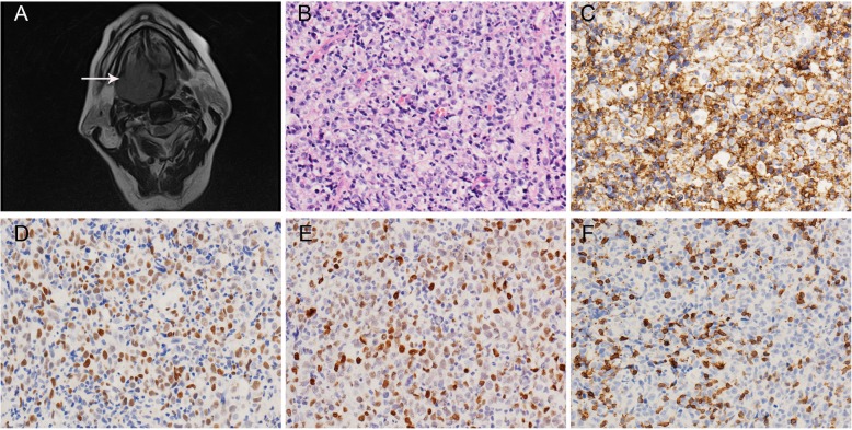
Results: All 7 lymphomas were localized at the base of the tongue. Six of the cases exhibited tongue base masses with smooth surface membranes. One case presented as multiple deep ulcers. The most common histologic subtype was diffuse large B-cell lymphoma (DLBCL), which occurred in five cases. The other two cases were mantle cell lymphoma (MCL) and peripheral T cell lymphoma, not otherwise specified (PTCL, NOS). One of the DLBCL cases was positive for HPV DNA and diffusely expressed P16 protein. During the follow up period, the MCL patient and an elderly DLBCL patient died. The remaining five patients were alive through the end of follow up.

Conclusions: Most lymphomas of the tongue base manifest as an endogenous mass without membranous change. The most common subtype of NHLs of the tongue base is DLBCL, and the occurrence at this site may have a good prognosis. With proper therapy, even late stage tongue base lymphomas can be suppressed and remain in remission.

Keywords: Diffuse large B-cell lymphoma; EBV; HPV; Mantle cell lymphoma; Non-Hodgkin’s lymphoma; Peripheral T cell lymphoma; Tongue base.


Conflict of interest statement

The authors declare that they have no competing interests.

Figures

Fig. 1
Fig. 1
Imaging and pathological findings of DLBCL (case 5). a. MRI showed a mass in the base of the tongue sticking to the pharyngeal cavity and making it obviously narrow. b. H&E showed immunoblastic large cells with an obvious nucleolus (200 x). c. Tumour cells diffusely expressed CD20 (200 x). d. Tumour cells were positive for C-myc (200 x). e. Tumour cells were positive for P53 (200 x). f. Tumour cells were negative for CD5 (200 x)
Fig. 2
Fig. 2
The case of DLBCL showing HPV DNA positivity (case 6). a. H&E showed a diffuse infiltrate of large cells with an obvious nucleolus and abundant cytoplasm (200 x). b. Tumour cells diffusely expressed CD20 (200 x). c. Immunohistochemistry staining showed diffuse and strong staining of P16 protein (40 x) d. Immunohistochemistry staining showed diffuse and strong staining of P16 protein (100 x). e. HPV DNA ISH showed brown punctate dots in the tumour cell nucleus or cytoplasm (400x).f. HPV RNA ISH all negative
Fig. 3
Fig. 3
Imaging and pathological findings of PTCL (case 3). a. CT showed a well-bordered cystic mass. b. H&E showed moderate to large cells with distorted nuclear contours (200 x). c. Tumour cell infiltrated squamous epithelium (400x). d. Tumour cells diffusely expressed CD3 (200x). e. Tumour cells were positive for CD4 (200x). f. Tumour cells were negative for CD8 (200x)
Fig. 4
Fig. 4
Imaging and pathological findings of MCL (case 2). a. CT showed an irregular soft tissue mass at the right posterior aspect of the tongue base. b. Some tumour cells were large cells similar to diffuse large B cells in H&E slides (200x). c. Some tumour cells were medium-sized with a clear cytoplasm (200x). d. Tumour cells were positive for CD5 (200x). e. Tumour cells were positive for Cyclin D1 (200x). f. Ki-67 staining of the tumour cells (200x)

References

    1. Lee JT, Paquette R, Sercarz JA, Wang MB. Mucosa-associated lymphoid tissue lymphoma of the lingual tonsil. Am J Otolaryngol. 2000;21:271–276. doi: 10.1053/ajot.2000.8382. - DOI - PubMed
    1. Lee YY, Van Tassel P, Nauert C, North LB, Jing BS. Lymphomas of the head and neck: CT findings at initial presentation. AJR Am J Roentgenol. 1987;149:575–581. doi: 10.2214/ajr.149.3.575. - DOI - PubMed
    1. Maheshwari GK, Baboo HA, Gopal U, Wadhwa MK. Primary extra-nodal non-Hodgkin's lymphoma of the cheek. J Postgrad Med. 2000;46:211–212. - PubMed
    1. Takahashi H, Fujita S, Okabe H, Tsuda N, Tezuka F. Immunophenotypic analysis of extranodal non-Hodgkin's lymphomas in the oral cavity. Pathol Res Pract. 1993;189:300–311. doi: 10.1016/S0344-0338(11)80514-5. - DOI - PubMed
    1. Singh T, Amirtham U, Satheesh CT, Sajeevan KV, Jain A, Lakshmaiah KC, Babu KG, Lokanatha D. Primary B cell non-Hodgkin's lymphoma of tongue. Indian J Cancer. 2010;47:84–86. doi: 10.4103/0019-509X.58873. - DOI - PubMed

Grants and funding