Review
. 2020 Oct;375(1):154-160.
doi: 10.1124/jpet.120.265058. Epub 2020 Apr 1.

Microglia and Other Myeloid Cells in Central Nervous System Health and Disease

Affiliations
Review

Microglia and Other Myeloid Cells in Central Nervous System Health and Disease

Adithya Gopinath et al. J Pharmacol Exp Ther. 2020 Oct.

Abstract

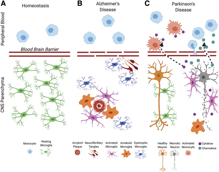
Mononuclear macrophages derived from the bone marrow (myeloid cells) are key cellular components of the innate immune system in different organs. In this minireview, we are focused on both brain and blood macrophages, known as microglia and monocytes, respectively. We provide a succinct summary of the cells' functions under both normal and pathologic conditions, with particular reference to common neurodegenerative disorders, such as Alzheimer and Parkinson disease. SIGNIFICANCE STATEMENT: In this minireview, we aim to summarize available literature on microglial and myeloid involvement in CNS disease, directing the reader toward relevant and translatable interpretations of myeloid cell function in CNS health and neurodegeneration.


Figures

Fig. 1.
Fig. 1.
Microglia at resting, activated, and dystrophic states are seen in MCAO (middle cerebral artery occlusion) unilateral stroke model brain tissue, accompanied by extensive peripheral myeloid cell infiltration. Within a coronal brain section from a mouse at 24 hours after the stroke in the contralateral hemisphere, (A) resting microglia are labeled with myeloid cell marker IBA1 (gray) and a marker of phagocytic and lysosomal function, CD68 (brown). (B) In the ipsilateral hemisphere, activated microglia at the edge of the lesion display a characteristic phenotype with increased IBA1 and CD68 expression, indicating active phagocytosis. (C) Dystrophic, fragmenting microglia can be observed within the stroke core. In addition to microglial activation, lesioned CNS tissue displays (D) extensive peripheral myeloid cell infiltration shown by Ly6B.2 immunostaining, specific to infiltrating peripheral myeloid cells. Identification of myeloid cell subtypes can be made using nuclear Nissl counterstain, as monocytes (mononuclear) or neutrophils (polymorphonuclear). Original magnification, 40× images. Scale bar, 20 μm.
Fig. 2.
Fig. 2.
Microglia and myeloid cell states in health and disease. While at homeostasis, (A) microglial activation in the CNS and alterations in peripheral blood myeloid cells are seldom seen, and dramatic changes in immune activation are seen in degenerative diseases such as AD and PD. (B) In AD, microglia become activated in response to amyloid formation and form phagocytic clusters attempting to remove this insoluble material. Neurofibrillary degeneration does not elicit microglial activation but coincides with microglial dystrophy; (C) in PD microglial activation, phagocytic activity and secretion of soluble signaling molecules are associated with response to dopaminergic neurons undergoing necroptosis in the midbrain. Secreted chemical factors also induce changes in peripheral myeloid populations, altering peripheral immune function and potentially inducing peripheral myeloid participation in dopaminergic degeneration.

References

    1. Alafuzoff I, Adolfsson R, Bucht G, Winblad B. (1983) Albumin and immunoglobulin in plasma and cerebrospinal fluid, and blood-cerebrospinal fluid barrier function in patients with dementia of Alzheimer type and multi-infarct dementia. J Neurol Sci 60:465–472. - PubMed
    1. Askew K, Li K, Olmos-Alonso A, Garcia-Moreno F, Liang Y, Richardson P, Tipton T, Chapman MA, Riecken K, Beccari S, et al. (2017) Coupled proliferation and apoptosis maintain the rapid turnover of microglia in the adult brain. Cell Rep 18:391–405. - PMC - PubMed
    1. Blum-Degen D, Müller T, Kuhn W, Gerlach M, Przuntek H, Riederer P. (1995) Interleukin-1 beta and interleukin-6 are elevated in the cerebrospinal fluid of Alzheimer’s and de novo Parkinson’s disease patients. Neurosci Lett 202:17–20. - PubMed
    1. Braak H, Del Tredici K. (2015) The preclinical phase of the pathological process underlying sporadic Alzheimer’s disease. Brain 138:2814–2833. - PubMed
    1. Braak H, Thal DR, Ghebremedhin E, Del Tredici K. (2011) Stages of the pathologic process in Alzheimer disease: age categories from 1 to 100 years. J Neuropathol Exp Neurol 70:960–969. - PubMed

Publication types

MeSH terms