. 2020 May 18;94(11):e00001-20.
doi: 10.1128/JVI.00001-20. Print 2020 May 18.

Identification of Reptarenaviruses, Hartmaniviruses, and a Novel Chuvirus in Captive Native Brazilian Boa Constrictors with Boid Inclusion Body Disease

Affiliations

Identification of Reptarenaviruses, Hartmaniviruses, and a Novel Chuvirus in Captive Native Brazilian Boa Constrictors with Boid Inclusion Body Disease

Fernando Froner Argenta et al. J Virol. .

Abstract

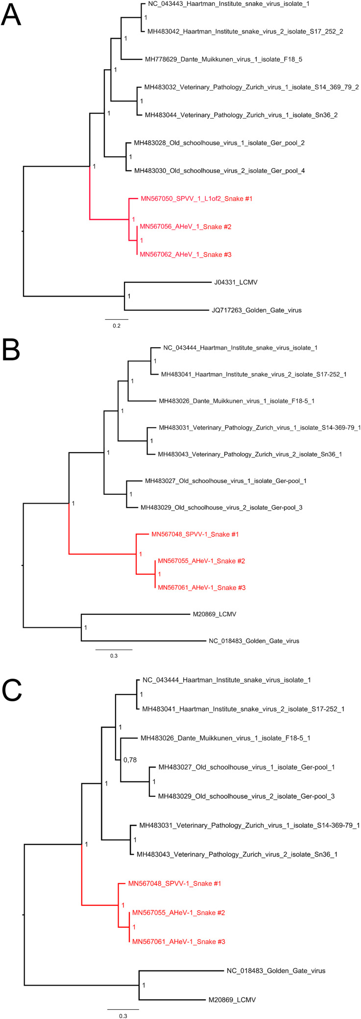
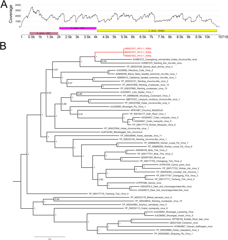
Boid inclusion body disease (BIBD) is a transmissible viral disease of captive snakes that causes severe losses in snake collections worldwide. It is caused by reptarenavirus infection, which can persist over several years without overt signs but is generally associated with the eventual death of the affected snakes. Thus far, reports have confirmed the existence of reptarenaviruses in captive snakes in North America, Europe, Asia, and Australia, but there is no evidence that it also occurs in wild snakes. BIBD affects boa species within the subfamily Boinae and pythons in the family Pythonidae, the habitats of which do not naturally overlap. Here, we studied Brazilian captive snakes with BIBD using a metatranscriptomic approach, and we report the identification of novel reptarenaviruses, hartmaniviruses, and a new species in the family Chuviridae The reptarenavirus L segments identified are divergent enough to represent six novel species, while we found only a single novel reptarenavirus S segment. Until now, hartmaniviruses had been identified only in European captive boas with BIBD, and the present results increase the number of known hartmaniviruses from four to six. The newly identified chuvirus showed 38.4%, 40.9%, and 48.1% amino acid identity to the nucleoprotein, glycoprotein, and RNA-dependent RNA polymerase, respectively, of its closest relative, Guangdong red-banded snake chuvirus-like virus. Although we cannot rule out the possibility that the found viruses originated from imported snakes, the results suggest that the viruses could circulate in indigenous snake populations.IMPORTANCE Boid inclusion body disease (BIBD), caused by reptarenavirus infection, affects captive snake populations worldwide, but the reservoir hosts of reptarenaviruses remain unknown. Here, we report the identification of novel reptarenaviruses, hartmaniviruses, and a chuvirus in captive Brazilian boas with BIBD. Three of the four snakes studied showed coinfection with all three viruses, and one of the snakes harbored three novel reptarenavirus L segments and one novel S segment. The samples originated from collections with Brazilian indigenous snakes only, which could indicate that these viruses circulate in wild snakes. The findings could further indicate that boid snakes are the natural reservoir of reptarena- and hartmaniviruses commonly found in captive snakes. The snakes infected with the novel chuvirus all suffered from BIBD; it is therefore not possible to comment on its potential pathogenicity and contribution to the observed changes in the present case material.

Keywords: arenavirus; boa constrictor; boid inclusion body disease; chuvirus; hartmanivirus; reptarenavirus.


Figures

FIG 1
FIG 1
Oral cavities of snakes with confirmed BIBD. (A) Animal 1, showing chronic ulcerative stomatitis. L, larynx. (B) Animal 3, showing focally ulcerated chronic stomatitis. Ch, choana.
FIG 2
FIG 2
Histological and immunohistological findings in the brain of animal 3 (A to C) and in the liver of animal 4 (E to G) and staining of tissues from animals without reptarena- or hartmanivirus infection (D and H). (A) Neurons exhibit the typical cytoplasmic eosinophilic IBs (arrowheads), which vary in size and can reach the size of and obscure the nucleus (arrows). HE stain. (B) Staining with the anti-pan-reptarenavirus antibody highlights the IBs depicted in the HE stain. Immunohistology, hemalaun counterstain. (C) Staining with the anti-pan-hartmani antibody highlights the IBs but also shows the presence of NP within the entire cytoplasm of infected cells (arrow and inset). Immunohistology, hemalaun counterstain. (D) Staining of reptrena- and hartmanivirus-negative tissue with anti-pan-reptarenavirus antibody. (E) Numerous hepatocytes exhibit a cytoplasmic eosinophilic IB of variable size (arrowheads). HE stain. (F) Staining with the anti-UHV NP antibody highlights individual IBs (arrowheads) and shows that some cells contain several small IBs (short arrow). Some larger IBs appear negative (large arrows). Immunohistology, hemalaun counterstain. (G) Staining with the anti-pan-reptarenavirus antibody shows the presence of abundant individual (arrowheads) and multiple (short arrows) IBs within hepatocytes. Again, a few larger IBs appear negative (large arrow). Immunohistology, hemalaun counterstain. (H) Staining of reptrena- and hartmanivirus-negative tissue with anti-pan-reptarenavirus antibody.
FIG 3
FIG 3
Maximum clade credibility tree of reptarenavirus L segments. The tree was constructed from amino acid sequences of the reptarenavirus representatives available from GenBank and those identified in this study, using the Bayesian MCMC method with the Jones model of amino acid substitution. Posterior probabilities are shown in each node.
FIG 4
FIG 4
Maximum clade credibility trees of reptarenavirus GPCs and NPs. (A) The phylogenetic tree based on the GPC amino acid sequences of the viruses identified in this study and those available in GenBank was constructed using the Bayesian MCMC method with the Blosum model of amino acid substitution. (B) The phylogenetic tree based on the NP amino acid sequences of the viruses identified in this study and those available in GenBank was constructed using the Bayesian MCMC method with the Jones model of amino acid substitution. The arrows indicate the S segments with the software-predicted recombination events listed in Table 5.
FIG 5
FIG 5
Maximum clade credibility trees for hartmanivirus RdRp, GPC, and NP. (A) The phylogenetic tree based on the RdRp amino acid sequences of the viruses identified in this study and those available in GenBank was constructed using the Bayesian MCMC method with the Blosum model of amino acid substitution. (B) The phylogenetic tree based on the GPC amino acid sequences of the viruses identified in this study and those available in GenBank was constructed using the Bayesian MCMC method with the Blosum model of amino acid substitution. (C) The phylogenetic tree based on the NP amino acid sequences of the viruses identified in this study and those available in GenBank was constructed using the Bayesian MCMC method with the Wag model of amino acid substitution.
FIG 6
FIG 6
Genome organization, similarity analyses, and phylogenetic tree of HFrV-1. (A) Genome organization and coverage (sequence from snake 1) of HFrV-1. The arrows represent the orientation of the open reading frames (ORFs). The L gene encodes RNA-dependent RNA polymerase (RdRp), the G gene encodes glycoprotein (GP), and the N gene encodes nucleoprotein (NP). The coverage (y axis) show the sequencing depth at each nucleotide position (x axis). (B) A maximum clade credibility tree based on the RdRp amino acid sequences of chuvirus-like viruses and chuviruses. The tree was constructed using the Bayesian MCMC method with the Blosum model of amino acid substitution. Posterior probabilities are shown in each node.

References

    1. Chang L, Jacobson ER. 2010. Inclusion body disease, a worldwide infectious disease of boid snakes: a review. J Exotic Pet Med 19:216–225. doi: 10.1053/j.jepm.2010.07.014. - DOI
    1. Schumacher J, Jacobson ER, Homer BL, Gaskin JM. 1994. Inclusion body disease in boid snakes. J Zoo Wildl Med 25:511–524.
    1. Stenglein MD, Sanders C, Kistler AL, Ruby JG, Franco JY, Reavill DR, Dunker F, Derisi JL. 2012. Identification, characterization, and in vitro culture of highly divergent arenaviruses from boa constrictors and annulated tree boas: candidate etiological agents for snake inclusion body disease. mBio 3:e00180-12. doi: 10.1128/mBio.00180-12. - DOI - PMC - PubMed
    1. Bodewes R, Kik MJ, Raj VS, Schapendonk CM, Haagmans BL, Smits SL, Osterhaus AD. 2013. Detection of novel divergent arenaviruses in boid snakes with inclusion body disease in The Netherlands. J Gen Virol 94:1206–1210. doi: 10.1099/vir.0.051995-0. - DOI - PubMed
    1. Hetzel U, Sironen T, Laurinmaki P, Liljeroos L, Patjas A, Henttonen H, Vaheri A, Artelt A, Kipar A, Butcher SJ, Vapalahti O, Hepojoki J. 2013. Isolation, identification, and characterization of novel arenaviruses, the etiological agents of boid inclusion body disease. J Virol 87:10918–10935. doi: 10.1128/JVI.01123-13. - DOI - PMC - PubMed

Publication types

LinkOut - more resources